Figure 1. Piezo2 expression in human iridocorneal angle tissues. A: Piezo2 (red) was expressed in the TM labeled with Collagen IV (ColIV) antibody (green), and the SC endothelial cells of
three human donors. The nuclei were stained with 4′,6-diamidino-2-phenylindole (DAPI in blue. B: Piezo2-positive cell fraction were 73.0% ± 19.1%, 39.9% ± 6.20%, and 22.3% ± 7.60% in the UM, CM, and JCT (n = 3). A statistically
significantly higher Piezo2-positive cell number was detected in the UM than in the JCT and the CM (p = 0.01, n = 3). No statistically
significant difference in fluorescence intensity was observed in different regions of the TM (n = 3). * p<0.05 with one-way
analysis of variance (ANOVA). C: Piezo2 expression was detected in HTM cells cultured in vitro with western blotting. β-actin was used as the reference protein.
D: Top panel: Piezo2 (red) were detected in NPE and PE cells, especially in NPE cells. Bottom panel: Piezo2 (red) was detected
in ciliary muscle robustly. In-focus images are shown in higher magnification on the bottom left. Scale bar: 100 µm. TM: trabecular
meshwork, SC: Schlemm’s canal, CB: ciliary body, UM: uveal meshwork, CM: corneoscleral meshwork, JCT: juxtacanalicular tissue,
AC: anterior chamber, PC: posterior chamber, NPE: non-pigmented epithelium cells, PE: pigmented epithelium cells, CM: ciliary
muscle.
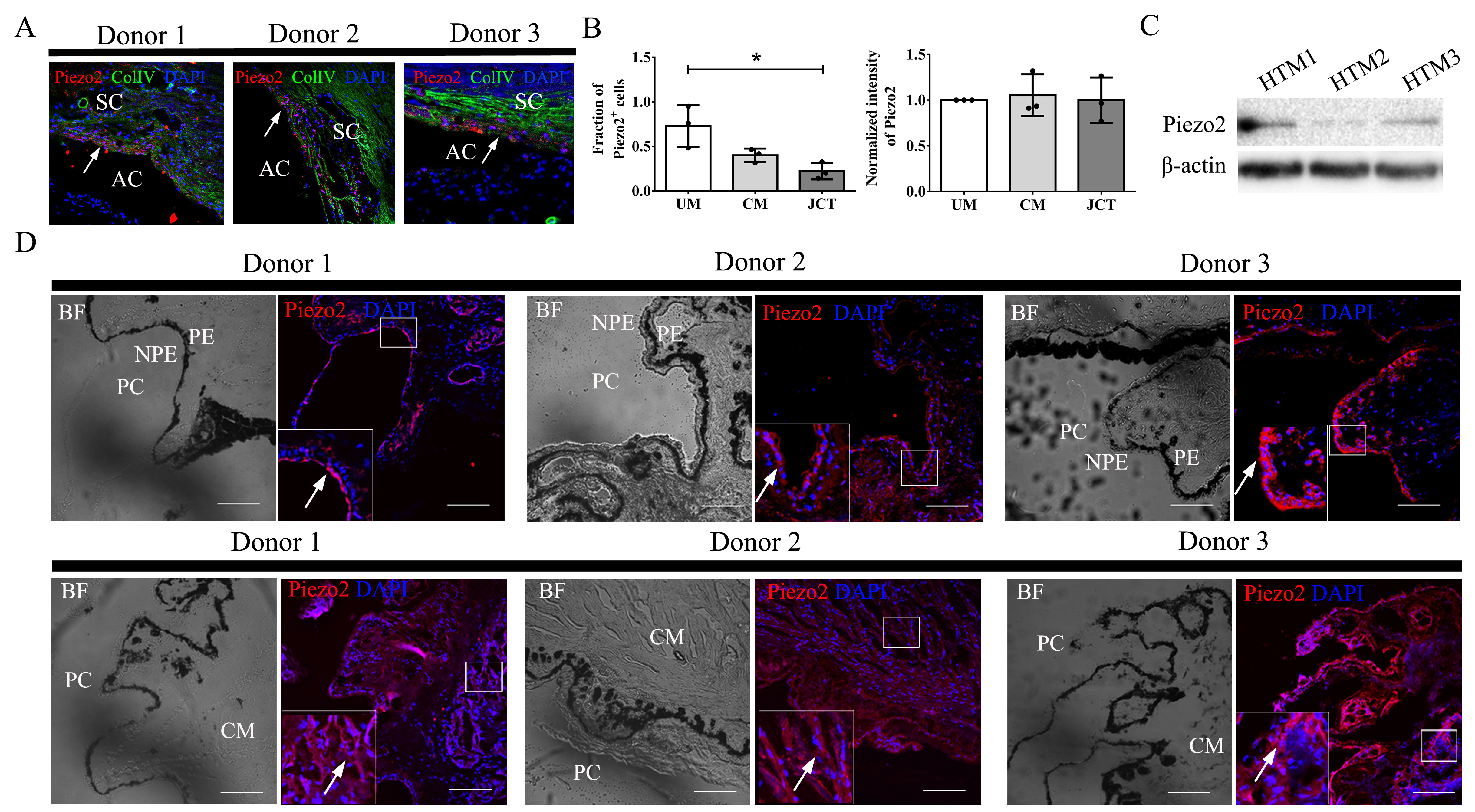
 Figure 1 of
Fang, Mol Vis 2020; 27:354-364.
Figure 1 of
Fang, Mol Vis 2020; 27:354-364.