Figure 4. In vivo studies of subretinal injection of cocultured cells in RCS rat animal models. A: Amplitudes of dark-adapted ten electroretinogram b-waves from each group of Royal College of Surgeon (RCS) rats at 4, 8,
12, and 20 weeks are shown (n=5 per group). A gradual decrease in amplitude is observed in all groups with no statistically
significant difference. Error bars indicate standard deviation, and asterisks (*) indicate statistical significance of p<0.05
(Mann–Whitney U test). B: Maximum cycle per degree, by which the RCS rats showed optokinetic responses at 4, 8, 12, and 20 weeks are shown (n=5 per
group). Preservation of optokinetic response was observed in group 3 (embryonic-stem-cell (ES)-derived RPE cells), group 4
(ES-derived photoreceptor precursor cells), and group 5 (1:1 mixture of both cells), compared to groups 1 (no injection) and
2 (media only injection), with statistical significance in some groups. Error bars indicate standard deviation, and asterisks
(*) indicate statistical significance of p<0.05 (Mann–Whitney U test). C: Immunofluorescent staining of the injected cells in the subretinal area in the RCS rats euthanized at 1 week and 8 weeks
were positive for anti-human nuclei (H. Mix) and the photoreceptor markers red and blue opsin.
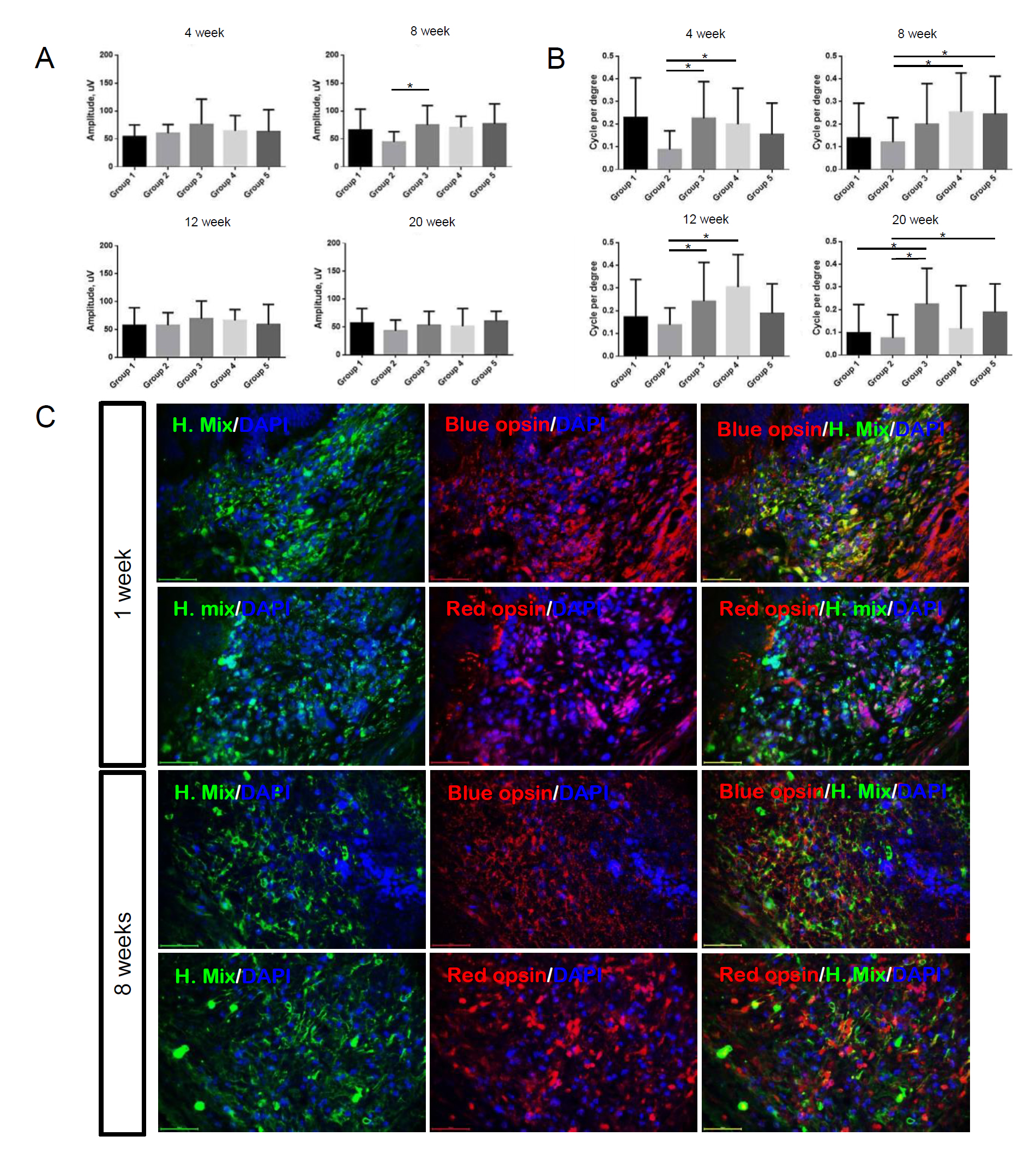
 Figure 4 of
Shin, Mol Vis 2021; 27:288-299.
Figure 4 of
Shin, Mol Vis 2021; 27:288-299.