Figure 4. Matrilin-2 immunohistochemistry. Control cornea: Mild immunopositivity in the epithelium, most prominent in the basal layer
(arrowheads) and moderate in the endothelium (inset). The stroma is immunonegative (A). BK cornea: The epithelial labelling is less pronounced and more evenly distributed (arrowheads) as compared to the control.
The subepithelial and middle part of the stroma is unstained, the pre-Descemet region, however, shows mild immunoreactivity
(arrows). The endothelium shows decreased staining intensity (inset) compared to the control (B). FECD cornea: The epithelium is thinner but does not show significant change in labelling intensity as compared to control.
Disease-specific vesicles are present between the epithelium and the Bowman membrane (stars). Mild matrilin-2 immunolabeling
appears throughout the stroma, which is more prominent in the pre-Descemet region (arrowheads). Unlike in BK, the endothelium
is stained sharply (C). HSV keratitis: The epithelium is irregular, the structure of the Bowman membrane is loose (arrows). There is delicate matrilin-2
expression in the subepithelial and middle part of the stroma, being more conspicuous in the pre-Descemet region (arrowheads).
A blood vessel (star) invades the stroma indicative of pathological neovascularization (D). Insets: The endothelium of the control cornea and corneas affected by the studied disorders. Scale bars refer to 100 μm
in the large figures and to 50 μm in the insets. Abbreviations: BK: bullous keratopathy; FECD: Fuchs' endothelial corneal
dystrophy; HSV: herpes simplex virus.
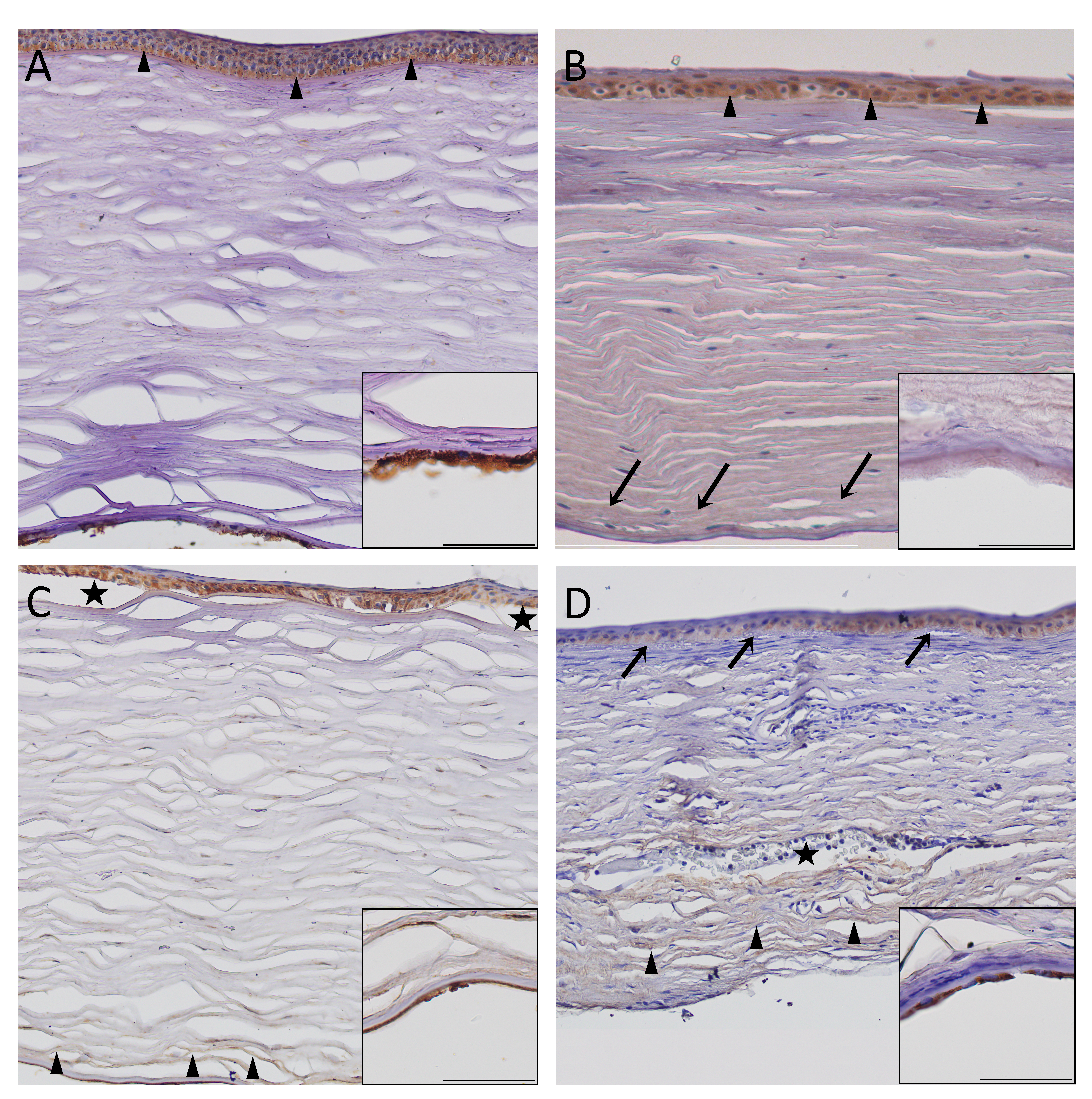
 Figure 4 of
Módis, Mol Vis 2021; 27:26-36.
Figure 4 of
Módis, Mol Vis 2021; 27:26-36.