Figure 3. Tenascin-C immunohistochemistry. Control cornea: The epithelium, particularly the basal layer, shows mild immunopositivity
(arrowheads). In addition, the endothelium (inset) shows a weak positivity regarding tenascin-C. Other components of the tissue
are unstained (A). BK cornea: Tenascin-C is expressed moderately in the epithelium. An intraepithelial vesicle (star) is forming, which is
characteristic of the disease. Bullae are present between the epithelium and Bowman layer (arrows). Intense expression appeared
throughout the stroma, most prominently in the pre-Descemet region (arrowheads). Endothelial signal intensity (inset) does
not differ significantly from the control (B). FECD cornea: In the basal cell layer of the epithelium intracellular oedema is detectable with increased tenascin-C expression
(arrows). A vesicle is present between the epithelium and Bowman layer (star). Tenascin-C expression is mild throughout the
stroma, however, with a gradual increase towards the pre-Descemet layer. The middle and the pre-Descemet region (arrowheads),
respectively, differ from the control at a high level of significance (p<0.001; C). HSV keratitis cornea: The epithelium displays cellular and nuclear pleiomorpism and disorganization of layering. In addition,
patchy loss of Bowman layer is revealed (arrow). Strong immunopositivity is present throughout the tissue, which is most prominent
in the epithelium. The endothelium (inset) expressed more intensely tenascin-C as compared to the other investigated conditions
(D). Insets: The endothelium of the control cornea and corneas affected by the studied disorders. Scale bars refer to 100 μm
in the large figures and to 50 μm in the insets. Abbreviations: BK: bullous keratopathy; FECD: Fuchs' endothelial corneal
dystrophy; HSV: herpes simplex virus.
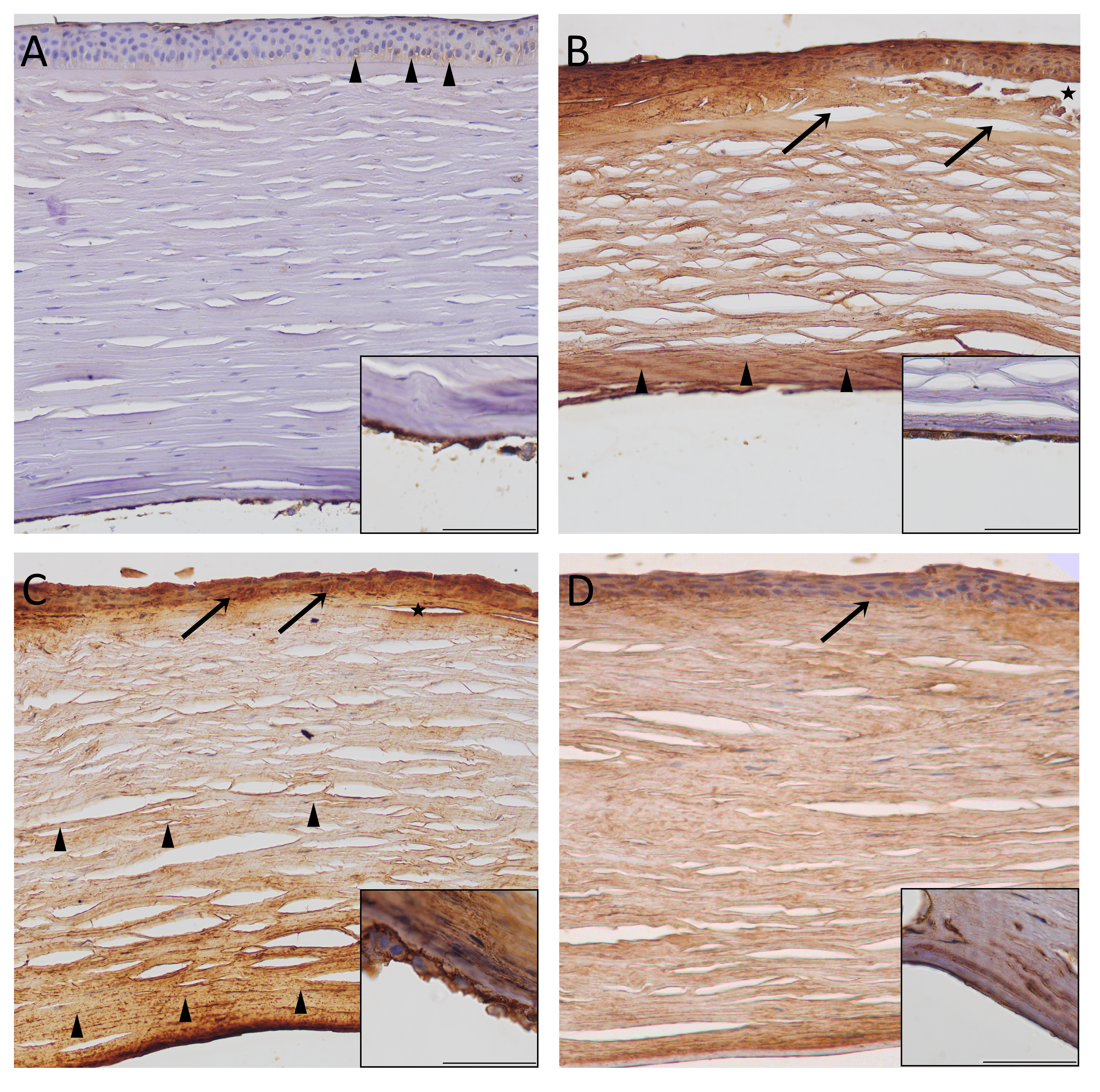
 Figure 3 of
Módis, Mol Vis 2021; 27:26-36.
Figure 3 of
Módis, Mol Vis 2021; 27:26-36.