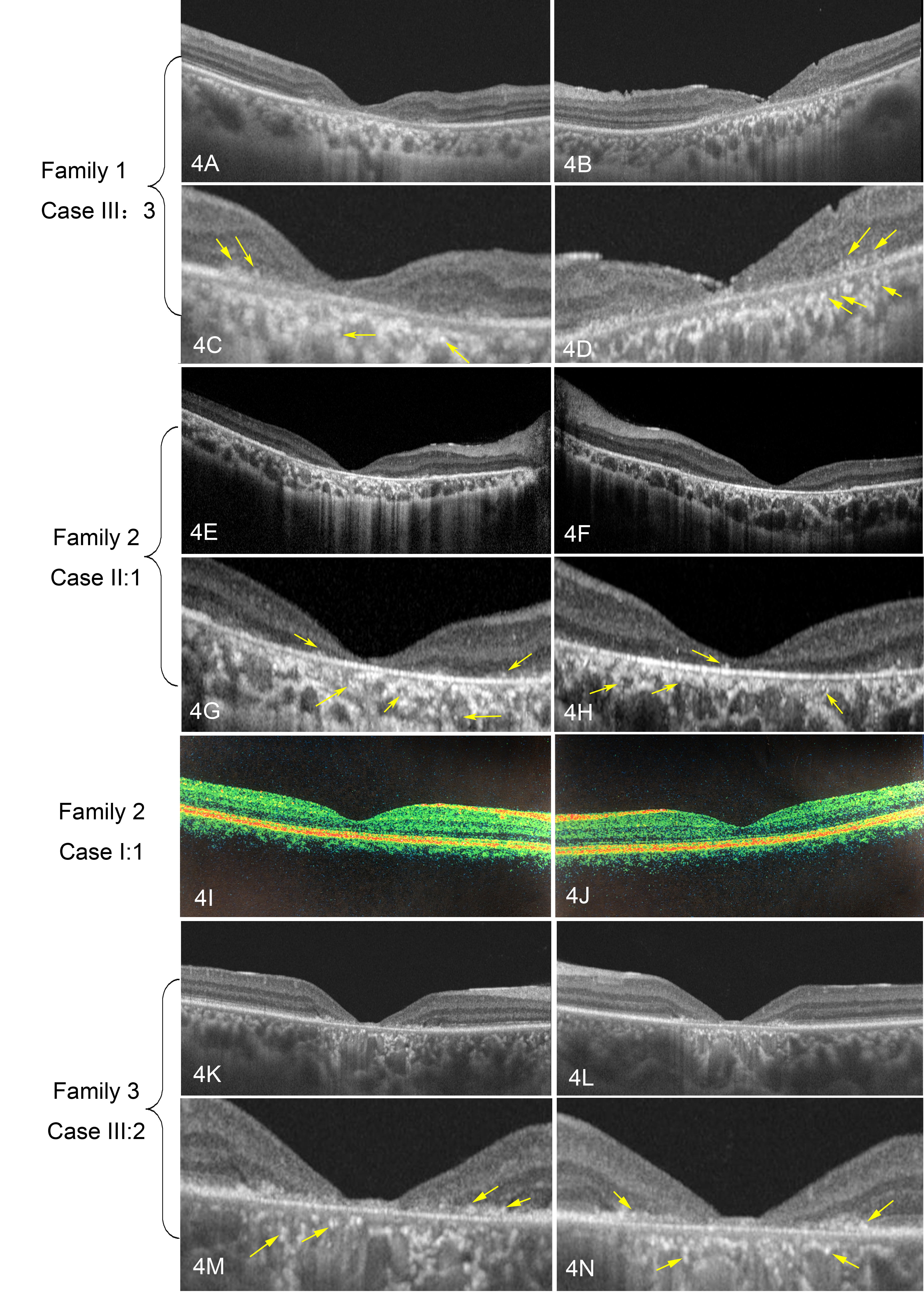
Figure 4. Optical coherence tomography (OCT) of patients with variants in AXTN7. Family 1 Case III:3 (a 54-year-old woman; A–D), Family 2 Case II:1 (an 18-year-old girl during the second visit; E–H), and Family 3 Case III:2 (a 32-year-old man; 4 K-4N) have a similar appearance. Optical coherence tomography (OCT) shows
thinning of the macula, with the outer layers and retinal pigment epithelium (RPE) affected more severely. The ellipsoid zone
disappears in the fovea area, indicating the appearance of cone-rod dystrophy (CORD). There are many intraretinal hyperreflective
dots, which are indicated by the yellow arrows. Panels C and D are enlargements of panels A and B, panels G and H are enlargements of panels E and F, and panels M and N are enlargements of panels K and L. Family 2 Case I:I (I and J) showed a normal OCT when he first consulted us at age 38.

 Figure 4 of
Zou, Mol Vis 2021; 27:221-232.
  Figure 4 of
Zou, Mol Vis 2021; 27:221-232.