Figure 7. Transmission electron microscopy pictures shows a direct connection between the microglia and the synapses. A: At day 1, microglia are found in the outer plexiform layer (OPL). B: The direct juxtaposition between the microglia and synaptic elements (arrow) is observed at day 3 (box). C: At day 14, microglia are found to extend their processes between sponge-like structures (star) in the OPL. M, microglia.
Scale bar: 5.0 μm for A, 0.5 μm for B and C.
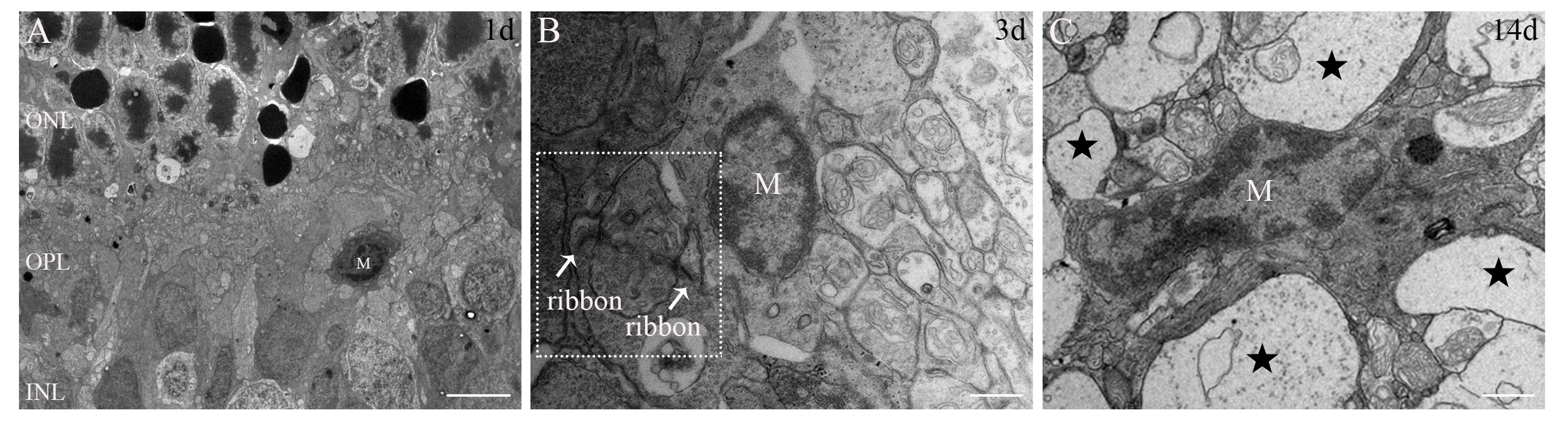
 Figure 7 of
Xu, Mol Vis 2021; 27:206-220.
Figure 7 of
Xu, Mol Vis 2021; 27:206-220.