Figure 5. Immunofluorescence against CD11b after light exposure. A: In the control retinas, ramified microglia are observed (arrow). B: One hour after light exposure, activated microglia migrate toward the outer retina. In addition, some microglia terminate
their processes in the outer plexiform layer (OPL; arrow). C: At day 3, some microglia are still terminating and stratifying their processes in the OPL (arrow). Some activated microglia
are observed in the subretinal space (arrowhead). D: Quantification of the number of microglia in the outer retina (including the OPL, outer nuclear layer, and subretinal space)
during retinal degeneration. n = 6. **p<0.01. Scale bar: 50 μm.
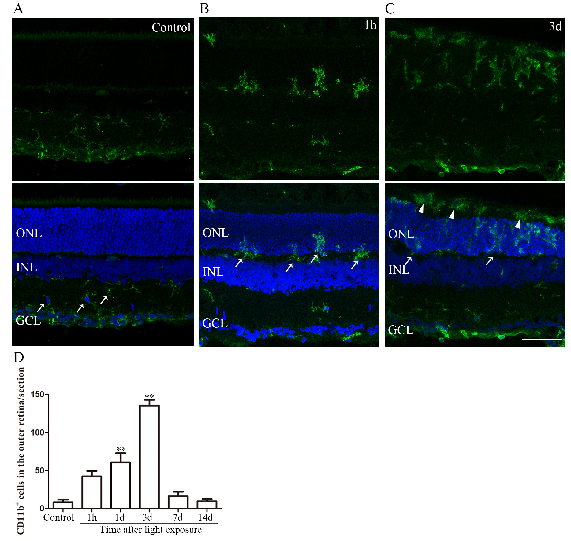
 Figure 5 of
Xu, Mol Vis 2021; 27:206-220.
Figure 5 of
Xu, Mol Vis 2021; 27:206-220.