Figure 3. Analysis of synaptic proteins of first synapses in the retina. A: Western blotting of postsynaptic density-95 (PSD-95). B: Western blotting of metabotropic glutamate receptor 6 (mGluR6). n = 4. *p<0.05, **p<0.01. C: In the control animals, mGluR6 immunoreactivity is observed in the outer plexiform layer. D–H: After light exposure, the staining is diminished during the early course of the degeneration. Scale bar: 50 μm.
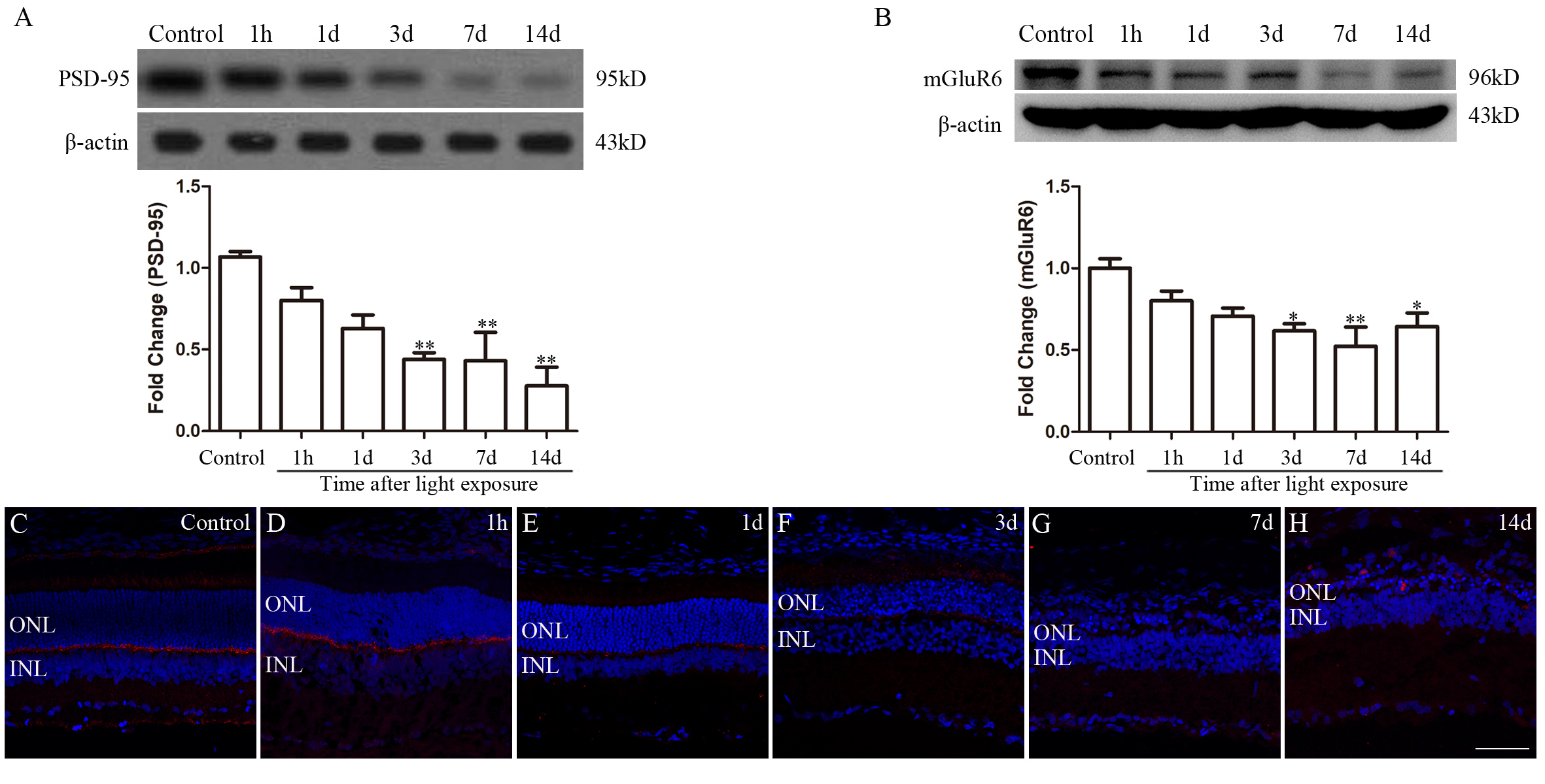
 Figure 3 of
Xu, Mol Vis 2021; 27:206-220.
Figure 3 of
Xu, Mol Vis 2021; 27:206-220.