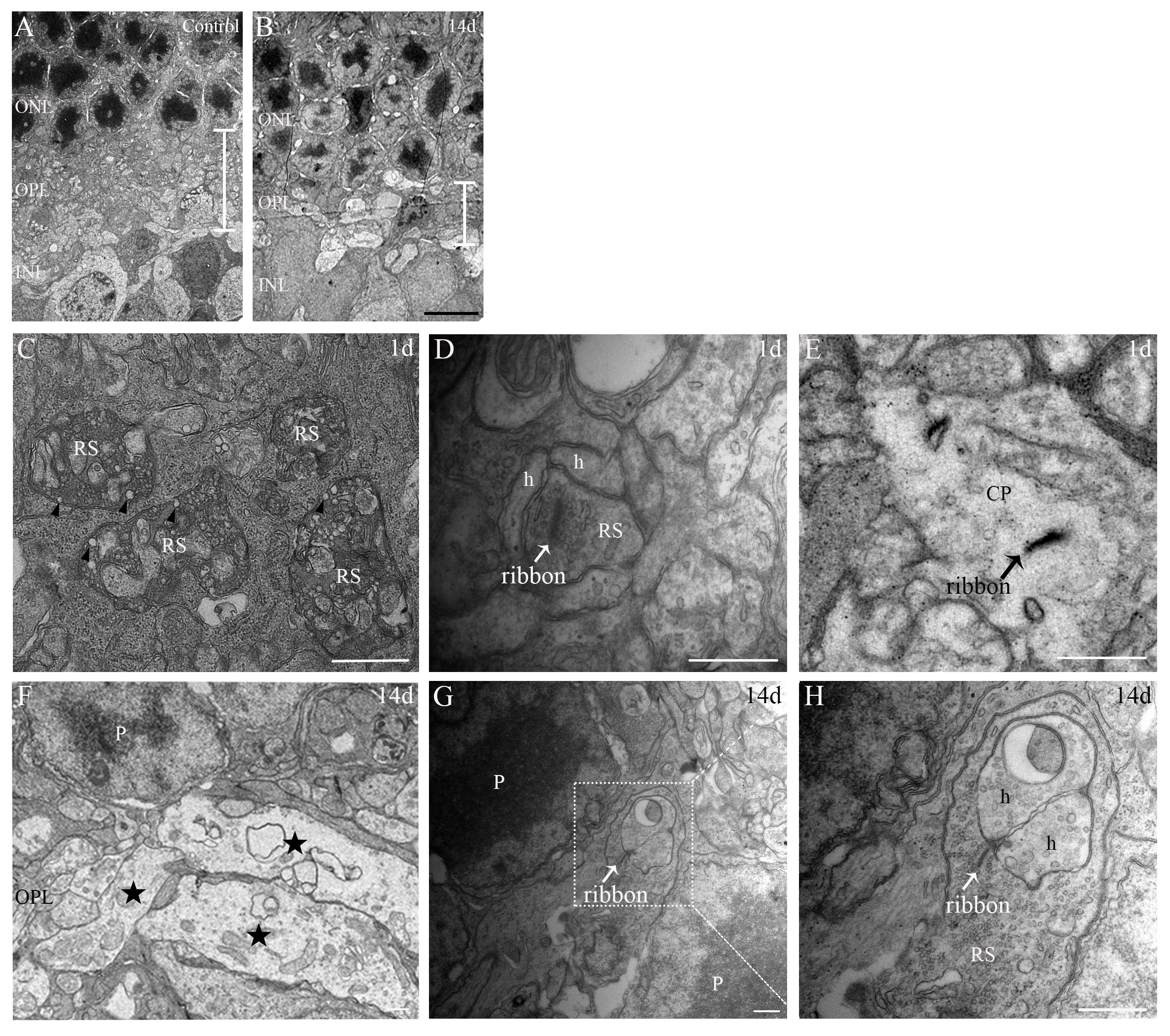
Figure 2. Transmission electron microscopy pictures of the retina show thinning of the OPL and degeneration of synapses in the OPL.
A, B: The outer plexiform layer (OPL) is thinner in the light exposed animals at day 14 after light exposure than in the control
animals. C: At day 1, many degenerated rod spherules (RSs) in the OPL have become very dark containing several vacuoles (arrowhead).
D, E: At day 1, presynaptic ribbons float in the cytoplasm of the RSs and cone pedicle (arrow). F: At day 14, the OPL is occupied by a sponge-like structure (star). G: At day 14, the RSs are observed to retract into the outer nuclear layer (ONL; box). H: Magnification of the box area in G. CP, cone pedicle; h, horizontal cell; P, photoreceptor; RS, rod spherule. Scale bar: 5.0 μm for A and B, 0.5 μm for C–H.

 Figure 2 of
Xu, Mol Vis 2021; 27:206-220.
  Figure 2 of
Xu, Mol Vis 2021; 27:206-220.