Figure 10. Immunoelectron microscopy shows engulfment of synaptic material by the microglia. A: In the control animals, microglia are observed in the inner part of the retina. Postsynaptic density-95 (PSD-95)-immunoreactive
electron-dense material is seldom seen inside the microglia. B, C: Magnification of the box area in A. No PSD-95-immunoreactive electron-dense material is observed in the cytoplasm of the microglia. D: A representative picture of a negative control at day 1 (without primary antibody). E, F: Magnification of the box area in D. No PSD-95-immunoreactive electron-dense material is observed in the cytoplasm of the microglia. G, H: At day 1, microglia are occasionally presented in the outer plexiform layer (OPL), and several 10 nm gold particles are
observed inside the microglia (arrow). H: Magnification of the box area in G. M, microglia; P, photoreceptor.
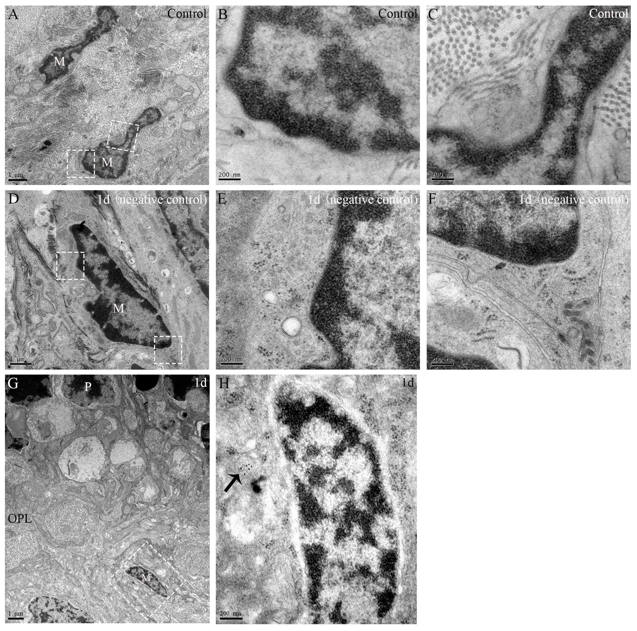
 Figure 10 of
Xu, Mol Vis 2021; 27:206-220.
Figure 10 of
Xu, Mol Vis 2021; 27:206-220.