Figure 3. Sub-cellular localization of WT TULP1 and P388S TULP1. Representative confocal microscopy images of human embryonic kidney
(HEK-293A) cells transfected with (A) green fluorescent protein (peGFP-C1), B: wild-type (WT) TULP1 enhanced GFP (eGFP), or (C) P388S TULP1 eGFP constructs (green) and stained with AlexaFluor 633 phalloidin (red). Scale bar = 50 μm. TULP1 eGFP images
are representative n≥5 biologic, independent replicates. Phalloidin images were representative of n≥3 separate independent
wells of a single transfection experiment.
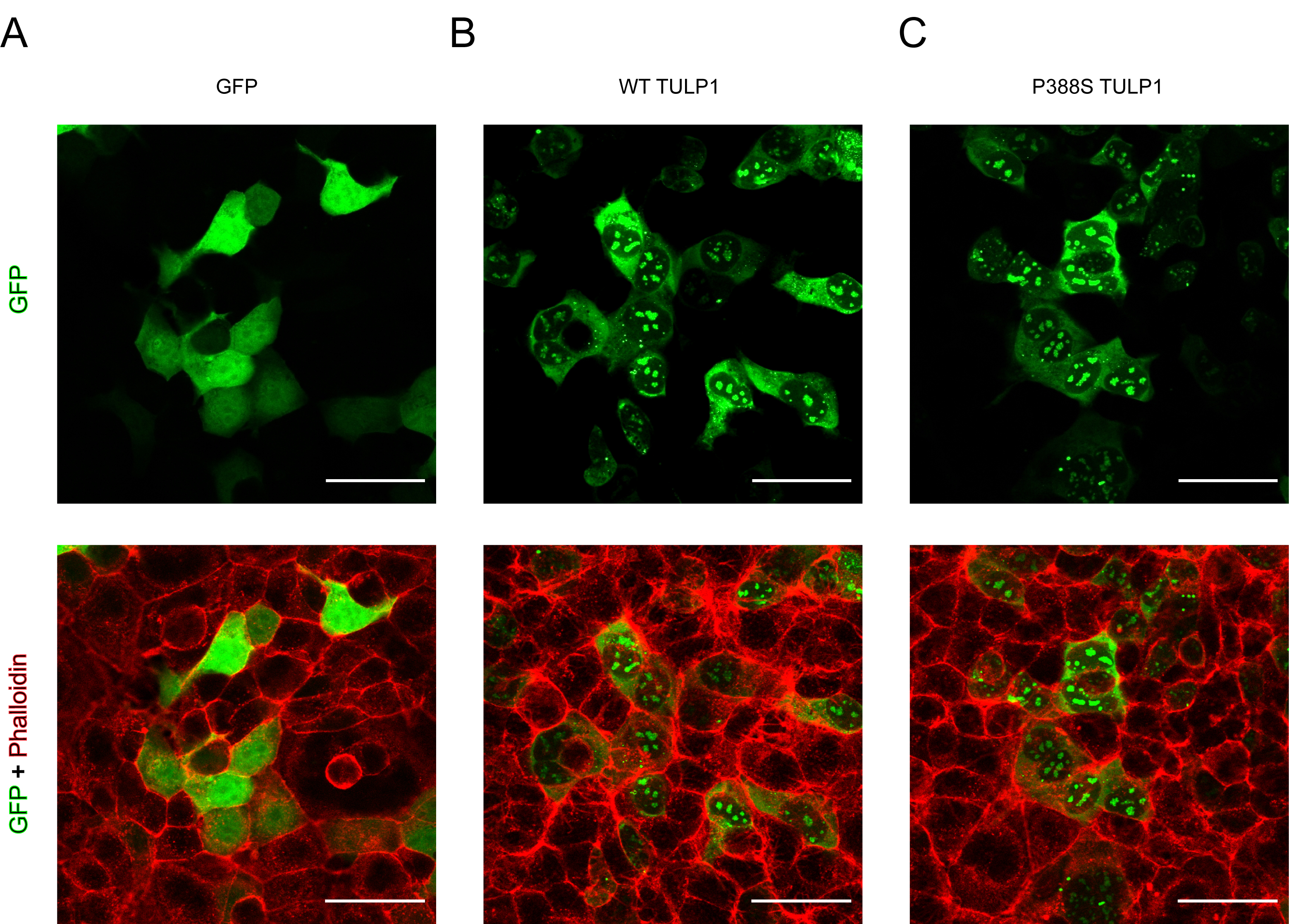
 Figure 3 of
Woodard, Mol Vis 2021; 27:179-190.
Figure 3 of
Woodard, Mol Vis 2021; 27:179-190.