Figure 2. Pedigree and in silico analysis of the pathogenic mutation. A: Pedigree of the consanguineous family with variant segregation based on Sanger sequencing. B: Multiple sequence alignment of TULP1 amino acid residues across species. Arrow indicates highlighted TULP1 residue. Alignments
were performed using Clustal Omega multiple sequence alignment software. C: In silico prediction findings related to the P388S mutation.
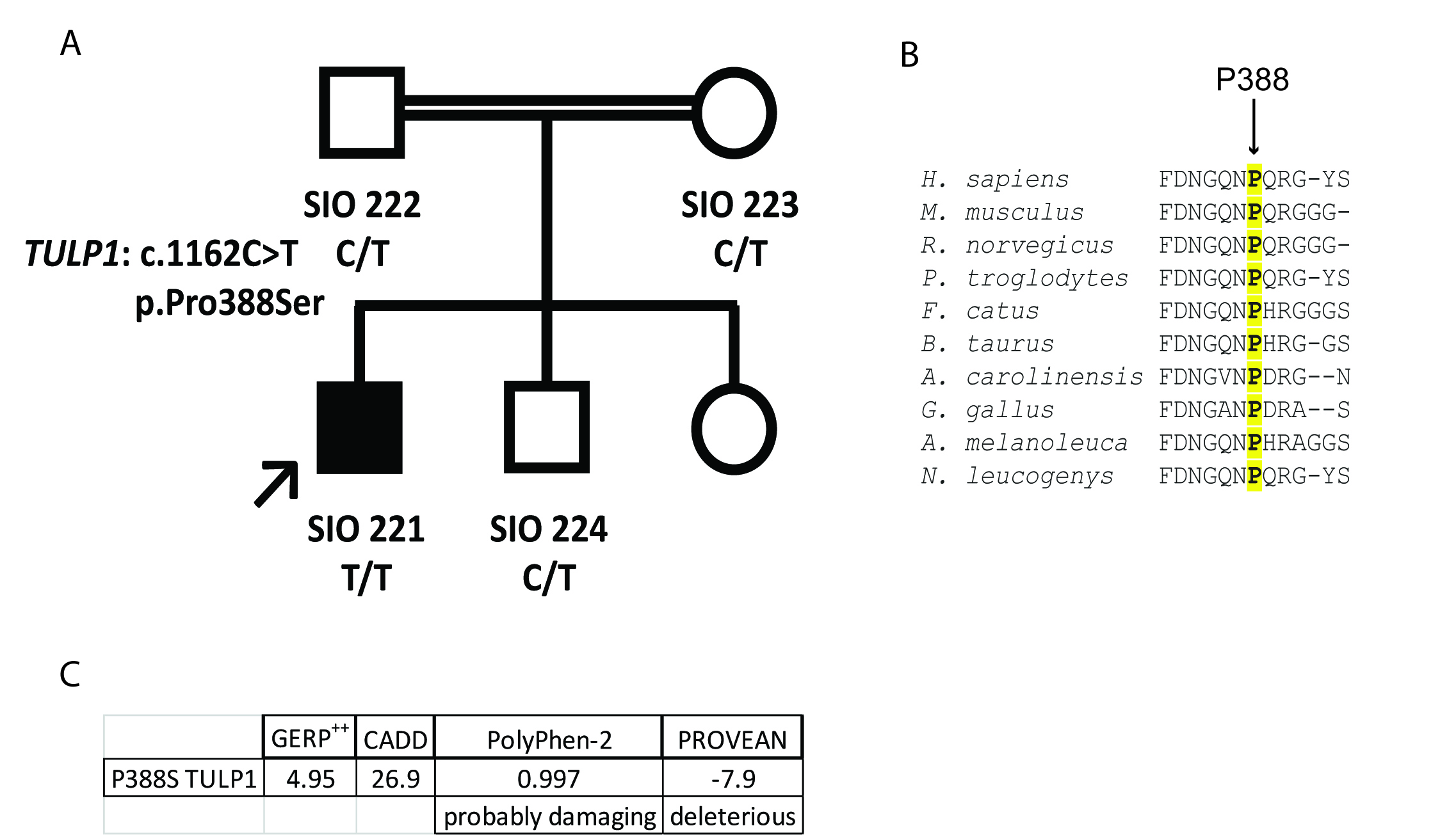
 Figure 2 of
Woodard, Mol Vis 2021; 27:179-190.
Figure 2 of
Woodard, Mol Vis 2021; 27:179-190.