Figure 1. Clinical characterization of the patient. A, B: Fundus photographs of the patient’s right and left eyes showing parafoveal RPE atrophy, bone spicule-like pigmentation,
and arteriolar attenuation. C, D:Fundus autofluorescence images showing parafoveal hypoautofluorescence corresponding to the area of RPE atrophy and a patchy
decrease in autofluorescence throughout the retina in both eyes. E, F: Optical coherence tomography (OCT) scans through the macula showing outer retinal atrophy with loss of the ellipsoid zone.
G–L: Fundus photographs, autofluorescence, and OCT images of an age-matched control subject.
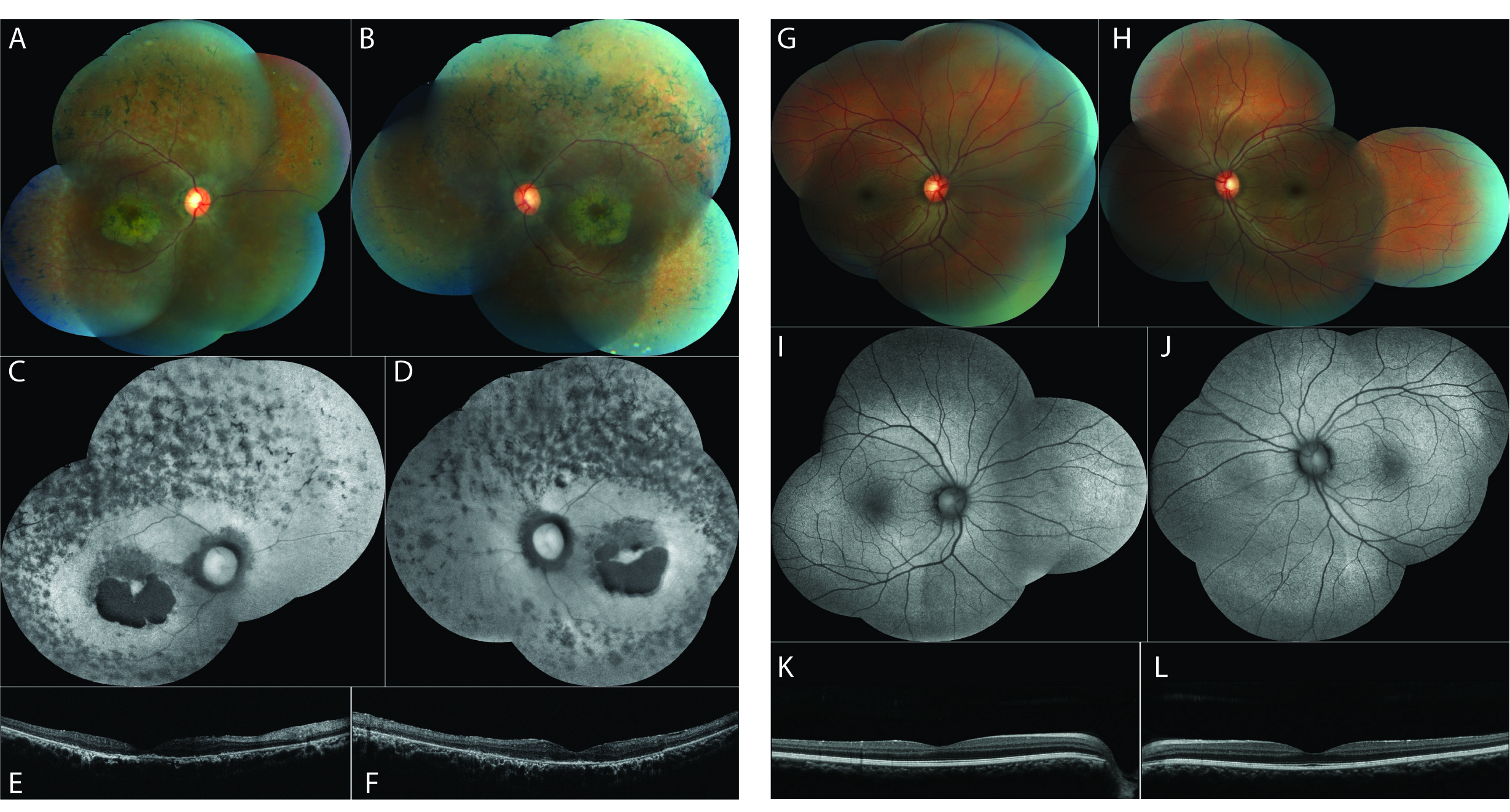
 Figure 1 of
Woodard, Mol Vis 2021; 27:179-190.
Figure 1 of
Woodard, Mol Vis 2021; 27:179-190.