Figure 5. Retinal imaging of patient RP3:03. A: Fundus imaging of patient RP3:03 shows a grayish-white region in the retinal periphery, a slightly pale papilla on the temporal
side, and progressively decreasing vessel sizes, as we move away from the papilla. B: Autofluorescence funduscopy discloses a discrete hyper-fluorescent ring surrounding the macular area and a large hypofluorescent
area in the peripheral retina. C: Macular optical coherence tomography (OCT) recording shows relative preservation of the foveal structure and the absence
of cystic space, but disorganization of the ellipsoid zone in the perifoveolar regions in both eyes, with abnormal epiretinal
membrane formation. RE: right eye; LE: left eye.
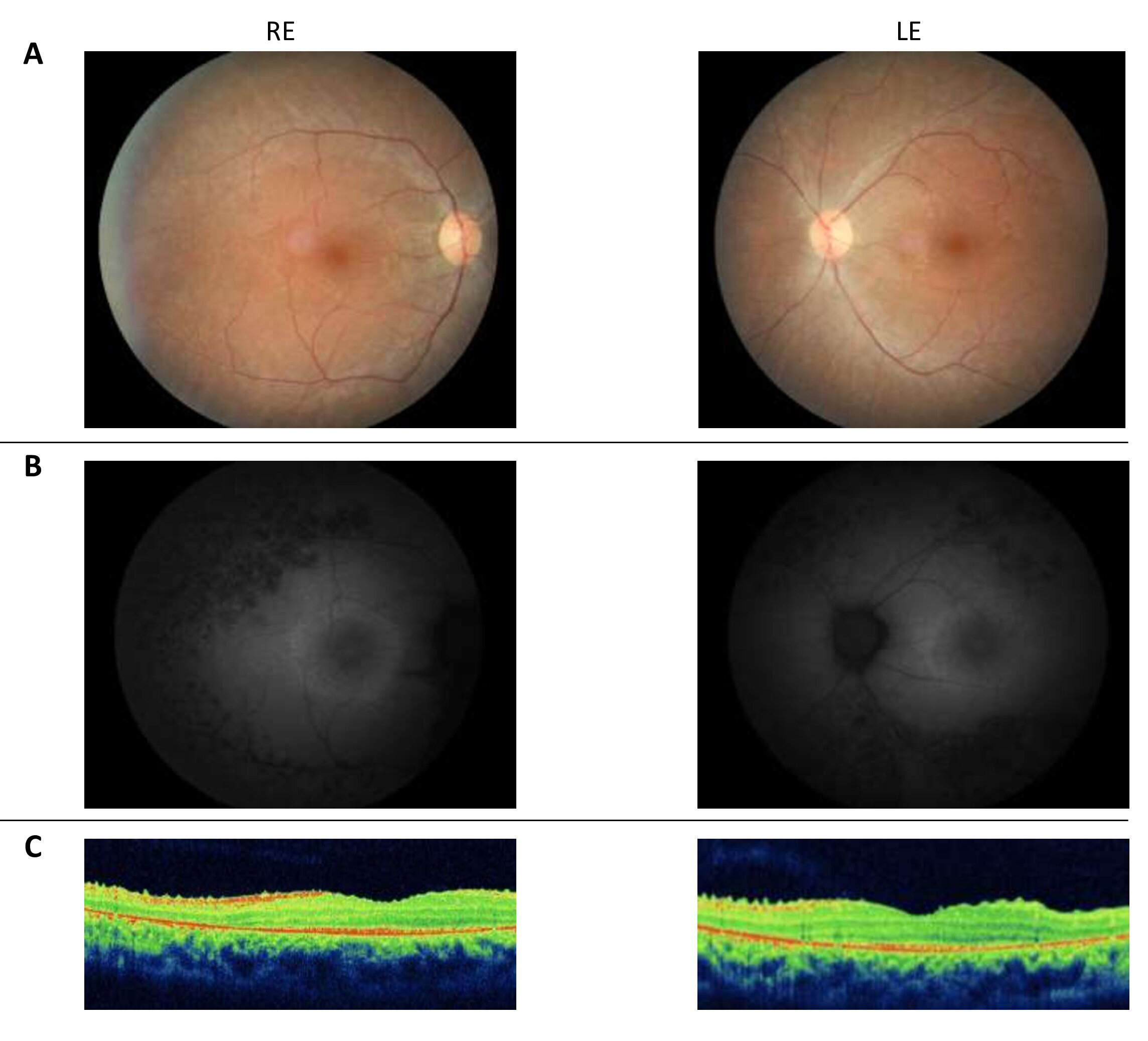
 Figure 5 of
Bouzidi, Mol Vis 2021; 27:17-25.
Figure 5 of
Bouzidi, Mol Vis 2021; 27:17-25.