Figure 4. Electroretinogram traces and visual field of patient RP3:03. A: Electroretinogram traces in the scotopic mode with blue light stimulation and the photopic mode with white and red lights,
and 30 Hz flickering light are unrecordable, showing the loss of rod and cone photoreceptors. B: The visual field of patient RP3:03 shows a severe impairment of the entire visual field with the preservation of tubular
vision in both eyes. RE: right eye; LE: left eye.
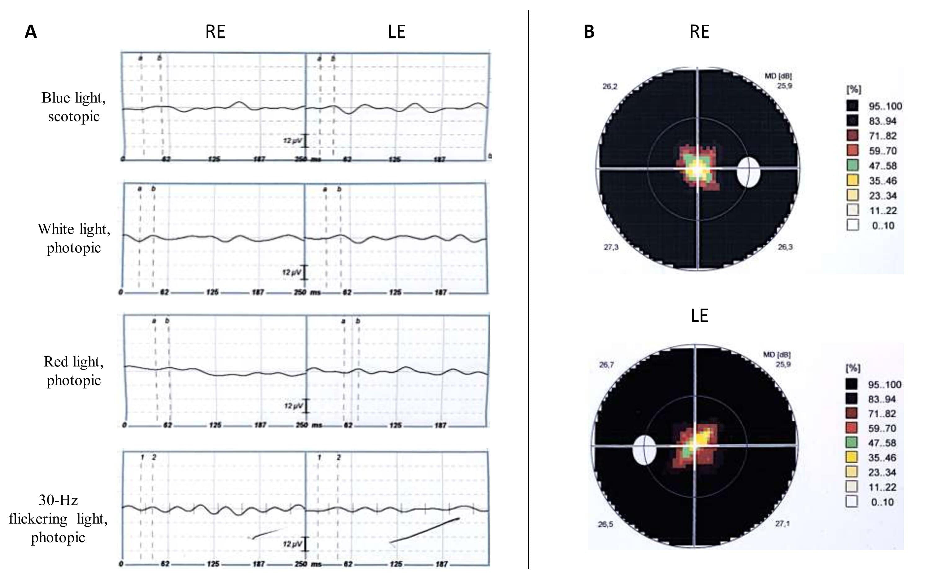
 Figure 4 of
Bouzidi, Mol Vis 2021; 27:17-25.
Figure 4 of
Bouzidi, Mol Vis 2021; 27:17-25.