Figure 3. Retinal autofluorescence and OCT imaging in patient RP2:03. A: Retinal autofluorescence examination of the right eye (RE) of patient RP2:03, presenting a large hypoautofluorescent central
macular area, marked by an arrow, and some hyper-autofluorescence in the inferior area of the optic nerve head. B: Macular optical coherence tomography (OCT) of the RE discloses cystoid macular edema and the loss of the ellipsoid zone
outside the fovea, reflecting rod photoreceptor loss.
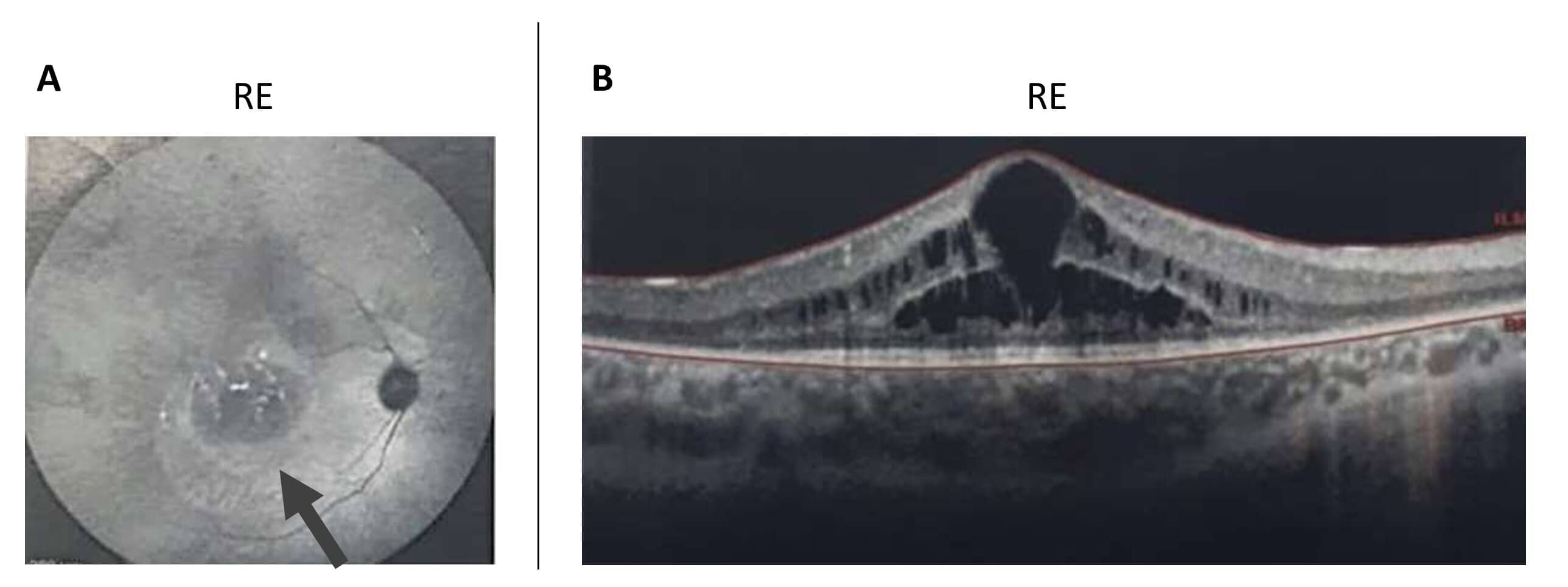
 Figure 3 of
Bouzidi, Mol Vis 2021; 27:17-25.
Figure 3 of
Bouzidi, Mol Vis 2021; 27:17-25.