Figure 1. Neuroprotective effects of SAHA in 661W photoreceptors. A: Two different HDAC6 inhibitors, one non-selective (Vorinostat; suberoylanilide hydroxamic acid, SAHA) and the other selective
(tubastatin A, TubA), were tested for their effect in protecting 661W photoreceptor cell survival and increasing NAD(P)H-dependent
cellular oxidoreductase enzyme activity using a colorimetric 3-(4,5-dimethylthiazol-2-yl)-2,5-diphenyltetrazolium bromide
(MTT) assay. Cell death was induced by the calcium ionophore A23187, selecting a dose that reduces the MTT assay readout by
50% (1 μM). The MTT readout could be improved upon in the presence of SAHA, but not tubastatin A over a 2 log unit dose range.
B: The same dose range of SAHA was tested using a high-resolution respirometric XF Seahorse assay examining the maximal respiratory
capacity (FCCP response). The oxygen consumption rate (OCR), a direct correlate of ATP reduction, demonstrated that application
of 3-isobutyl-1-methyl-xanthine (IBMX) reduced the OCR by about 40%, whereas SAHA was able to prevent that decline over the
same dose range as in (A). C: IBMX (600 μM) was shown to induce fragmentation in 661W cells stained with 10-N-nonyl acridine orange (NAO), an acridine
orange derivative that marks the inner mitochondrial membrane in whole cells. SAHA treatment prevented mitochondrial fission,
resulting in a fused phenotype. D: Cell extracts of 661W cells were probed for acetylated tubulin (AcTub) and histones (AcHH3 and 4) using western blotting.
Tubulin and glyceraldehyde 3-phosphate dehydrogenase (GADPH) were provided as controls. The typical signature for HDAC6 was
elucidated, stress increased tubulin and histone acetylation, but only tubulin acetylation, and not histone acetylation, was
further increased by SAHA. Data are shown as mean ± standard deviation (SD; n = 3–12), or representative images are shown.
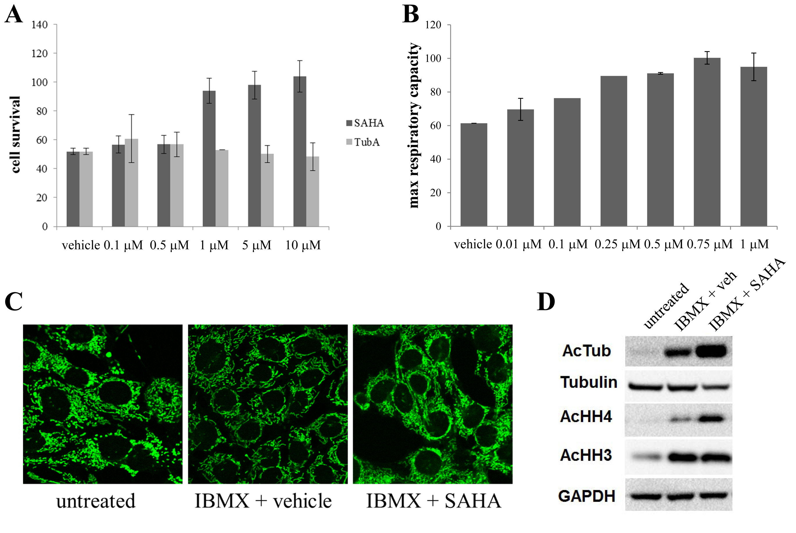
 Figure 1 of
Perron, Mol Vis 2021; 27:151-160.
Figure 1 of
Perron, Mol Vis 2021; 27:151-160.