Figure 8. Light micrographs of retinal cross-section (H&E staining). Hematoxylin and eosin (H&E) staining was performed for rabbits
with an intravitreal injection of inhibitor of hyaluronic acid-Vibrio mimicus collagenase that remains active (iHA-VMA) or VMA, post-euthanasia. The retinal morphology of the eyes treated with iHA-VMA
(A, B) was similar to that of the control eyes. Eyes injected with VMA (C, D) show intracellular edema, a significant increase in layer thickness, merging of the ONL and the INL (marked with asterisks
in D), or rarification in the retinal layers and inflammatory cells above the retina (marked with arrows in C), possibly due to vascular leakage. The corresponding contralateral, control eyes (C, D) show no sign of toxicity. In each panel, the control and treated eyes belong to the same rabbit. GCL, ganglion cell layer;
IPL, inner plexiform layer; INL, inner nuclear layer; ONL, outer nuclear layer; PL, photoreceptor layer. Scale bar = 50 µm.
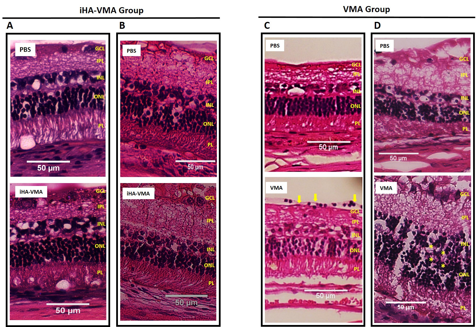
 Figure 8 of
Santra, Mol Vis 2021; 27:125-141.
Figure 8 of
Santra, Mol Vis 2021; 27:125-141.