Figure 2. Ophthalmic imaging apparatus setup using mice and lumpfish. A: Mouse on the Bioptigen spectral domain optical coherence tomography (SD-OCT) rodent stage showing the positions of the three-dimensional
(3D)-printed parts and the 78 diopter (D) lens. B: FiLMiC Pro imaging of mouse fundus on an iPod screen. C: Ocular structures of lumpfish being imaged manually at tank side. D: FiLMiC Pro imaging of lumpfish fundus on an iPod screen.
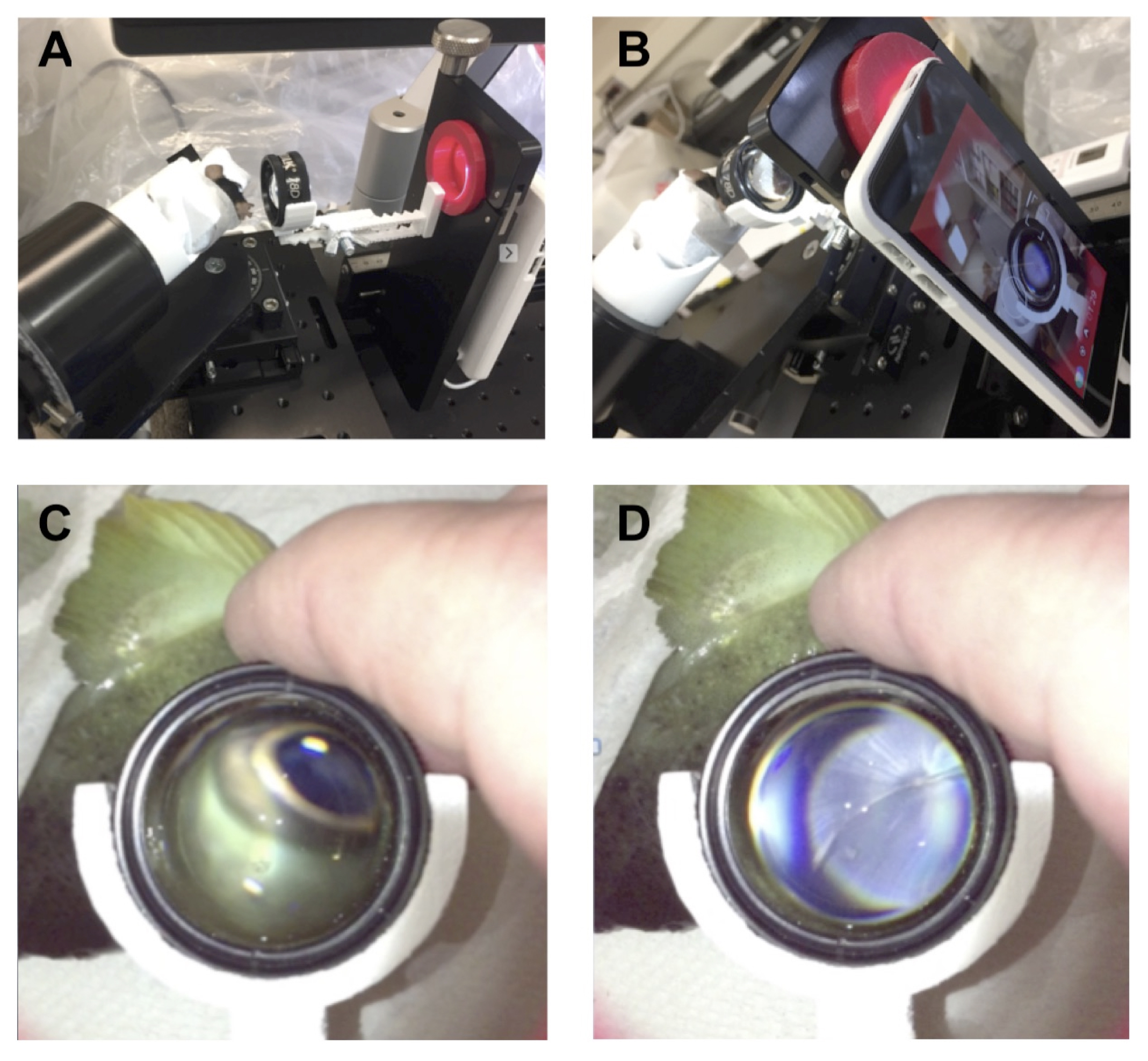
 Figure 2 of
McDonald, Mol Vis 2021; 27:117-124.
Figure 2 of
McDonald, Mol Vis 2021; 27:117-124.