Figure 2. Imaging of patients MOL0931–1 and MOL0931–2. A–D: Optical coherence tomography (OCT) map and representative macular scans of MOL0931–1 at the age of 18 years, before treatment
(A,C) and following treatment with 125 mg acetazolamide three times daily (B,D). E–H: Goldmann visual fields of MOL0931–1 at age 18 (E,F) and his sister MOL0931–2 at age 15 (G,H). I–J: OCT scans of MOL0931–2 at age 22.
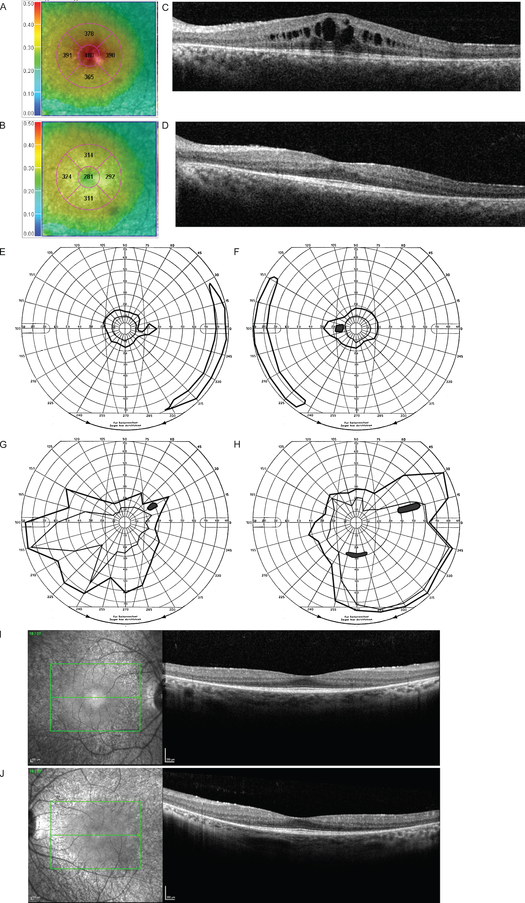
 Figure 2 of
Ruberto, Mol Vis 2021; 27:107-116.
Figure 2 of
Ruberto, Mol Vis 2021; 27:107-116.