Figure 3. Cogeneration of the neural retina and pigmented epithelium is enhanced by the scraping differentiation method. A: Proportion of retinal organoids (ROs) associated with pigmented epithelium domain. The bar charts summarized data from three
independent experiments using four different human pluripotent stem cell (hPSC) lines and presented as mean ± standard deviation.
*p<0.05. B: Pigmented epithelium (PE) of pigmented domain shown by EZRIN (green, upper), MERTK (red, upper), and PMEL17 (green, lower).
C: Schematic representation of CHX10, MITF, and AQP-1 staining in ROs obtained with the dissection and scraping methods. D: Immunostaining of retinal progenitor cell marker CHX10, ciliary epithelium marker AQP-1, and PE marker MITF. Nuclei were
stained with 4′,6-diamidino-2-phenylindole (DAPI, blue). Arrowheads indicate relevant staining of each marker.
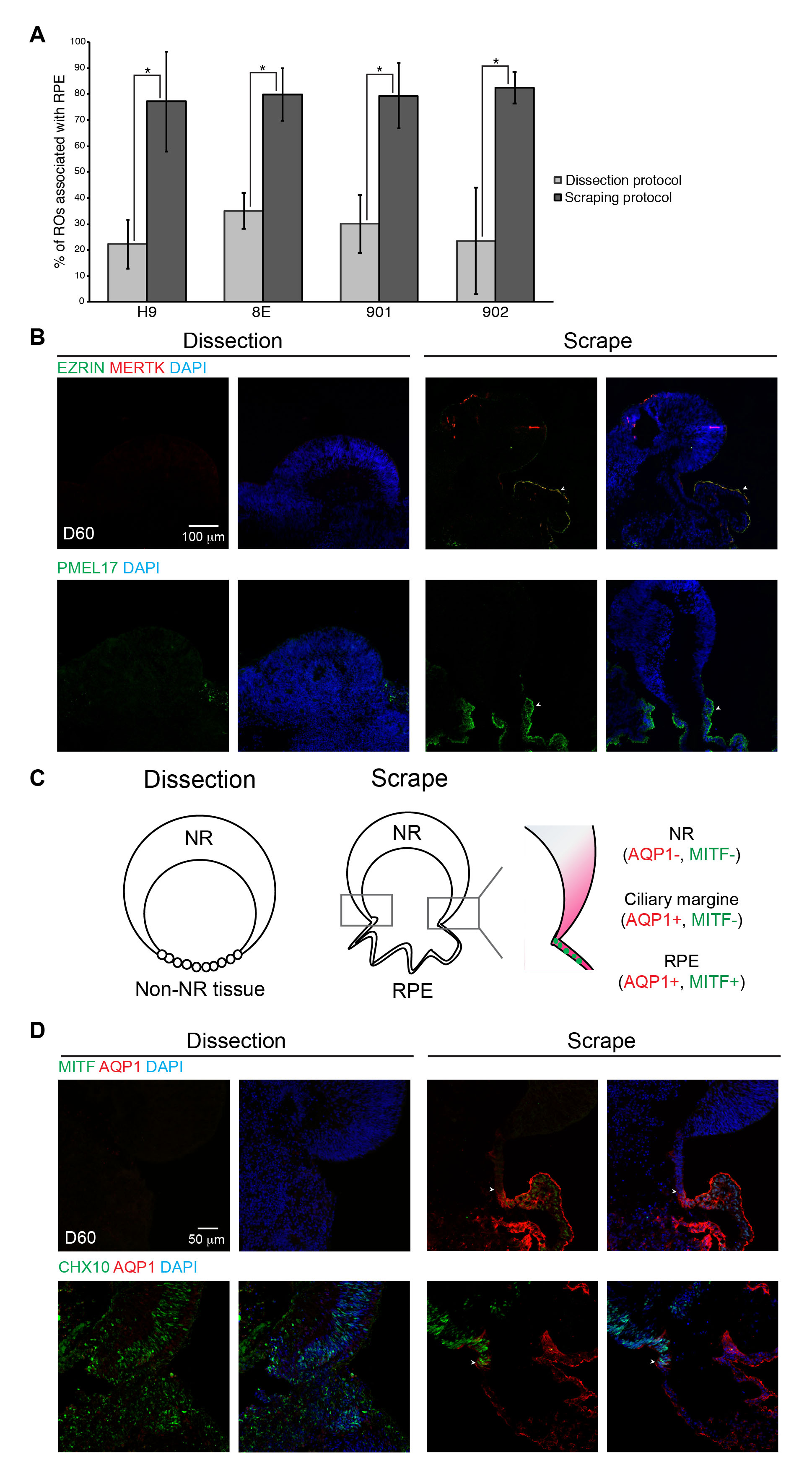
 Figure 3 of
Regent, Mol Vis 2020; 26:97-105.
Figure 3 of
Regent, Mol Vis 2020; 26:97-105.