Figure 2. The scraping method improves the yield of ROs with morphology similar to those obtained with dissection. A: Quantification of the number of retinal organoids (ROs) produced by the scraping and dissection methods, using human pluripotent
stem cells (hPSCs) from one well of a six-well plate. The bar charts summarized data from three independent experiments using
four different hPSC lines and presented as mean ± standard deviation. *p<0.05; n.s., non-significant. B: Immunohistochemistry analysis of H9 human embryonic stem cell (hESC)-derived ROs using antibodies against markers for retinal
ganglion cells (BRN3A, green), horizonal or amacrine cells (CALB, red), photoreceptor progenitor cells (RCVRN, red), Müller
glia (CRALBP, green), retinal progenitor cells or bipolar cells (CHX10, green), rod bipolar cells (PKCα, red) and photoreceptors
(RHO, green), S-cones (OPN1SW, yellow), and L/M-cones (OPNL/MW, red). C: Ciliary markers showing the basal bodies (PCTN, red, left panel) and the cilia (ARL13B, green, right panel) of photoreceptors.
Nuclei were stained with 4′,6-diamidino-2-phenylindole (DAPI, blue). Arrowheads indicate relevant staining of each marker.
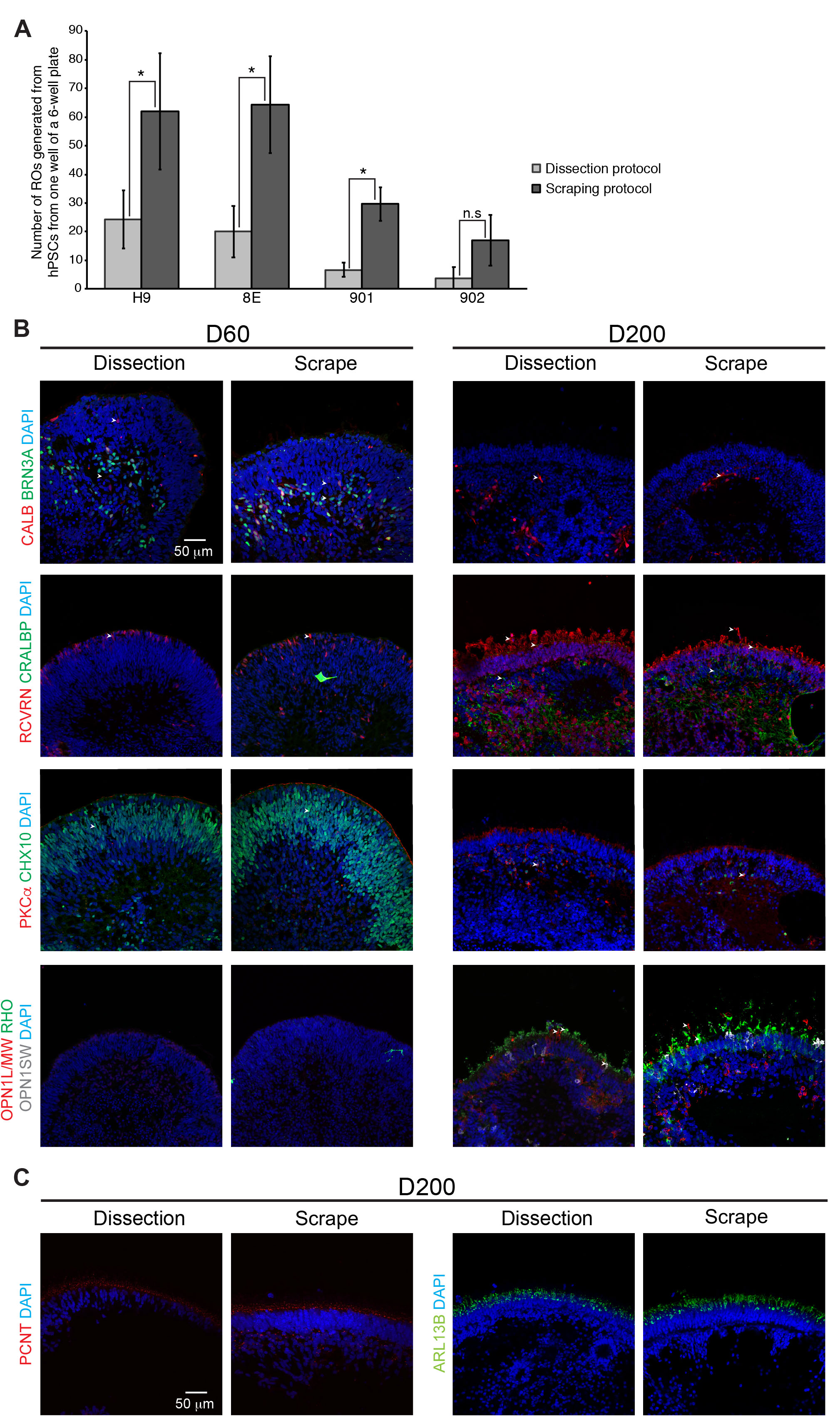
 Figure 2 of
Regent, Mol Vis 2020; 26:97-105.
Figure 2 of
Regent, Mol Vis 2020; 26:97-105.