Figure 1. Description of the scraping protocol for differentiating retinal organoids. A: Schematic representation of the scraping differentiation protocol used in this study. hPSCs: human pluripotent stem cells;
EBs: embryoid bodies; ROs: retinal organoids; NIM: neural induction medium; RIM: retinal induction medium; IGF1: insulin-like
growth factor 1. B:Representative bright-field images of EBs, cell clumps generated by the scraping method, and differentiating organoids (from
D30 to D200). C: Representative bright-field images of differentiating ROs 10 days after scraping from hESC line H9 and hiPSC lines 1C, 2D,
8E, 901, and 902. White asterisks: optic vesicle-like structures; white arrowheads: neural retina; black arrowheads: pigmented
epithelium domains; black arrow: presumptive photoreceptor cilia.
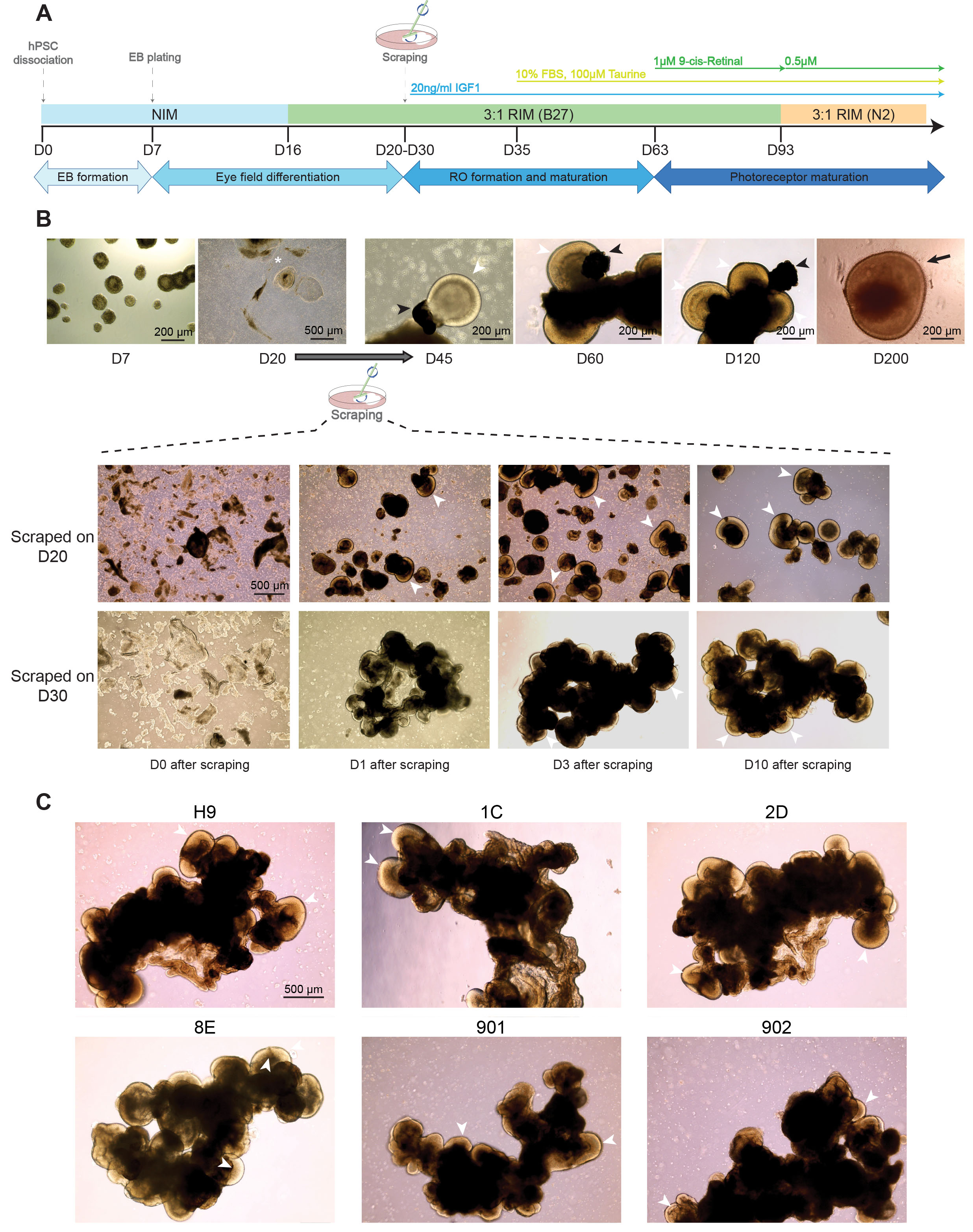
 Figure 1 of
Regent, Mol Vis 2020; 26:97-105.
Figure 1 of
Regent, Mol Vis 2020; 26:97-105.