Figure 5. Crystal violet staining. Cell proliferation was assessed after 6 days of treatment with 10 ng/ml basic fibroblast growth factor
(bFGF), 5 ng/ml transforming growth factor beta 1 (TGF-β), 10 ng/ml platelet-derived growth factor subunit-BB (PDGF-BB), 200
µg/ml ascorbic acid (AsA), 1 mg/ml dexamethasone (DEX), and 5 µM H2O2. Measurements were performed in triplicate; data were represented as % versus untreated (means ± standard deviation, SD).
The analysis of variance (ANOVA) with Tukey’s post-hoc correction was performed. *p<0.001 versus untreated (NT) primary cultured
hyalocytes.
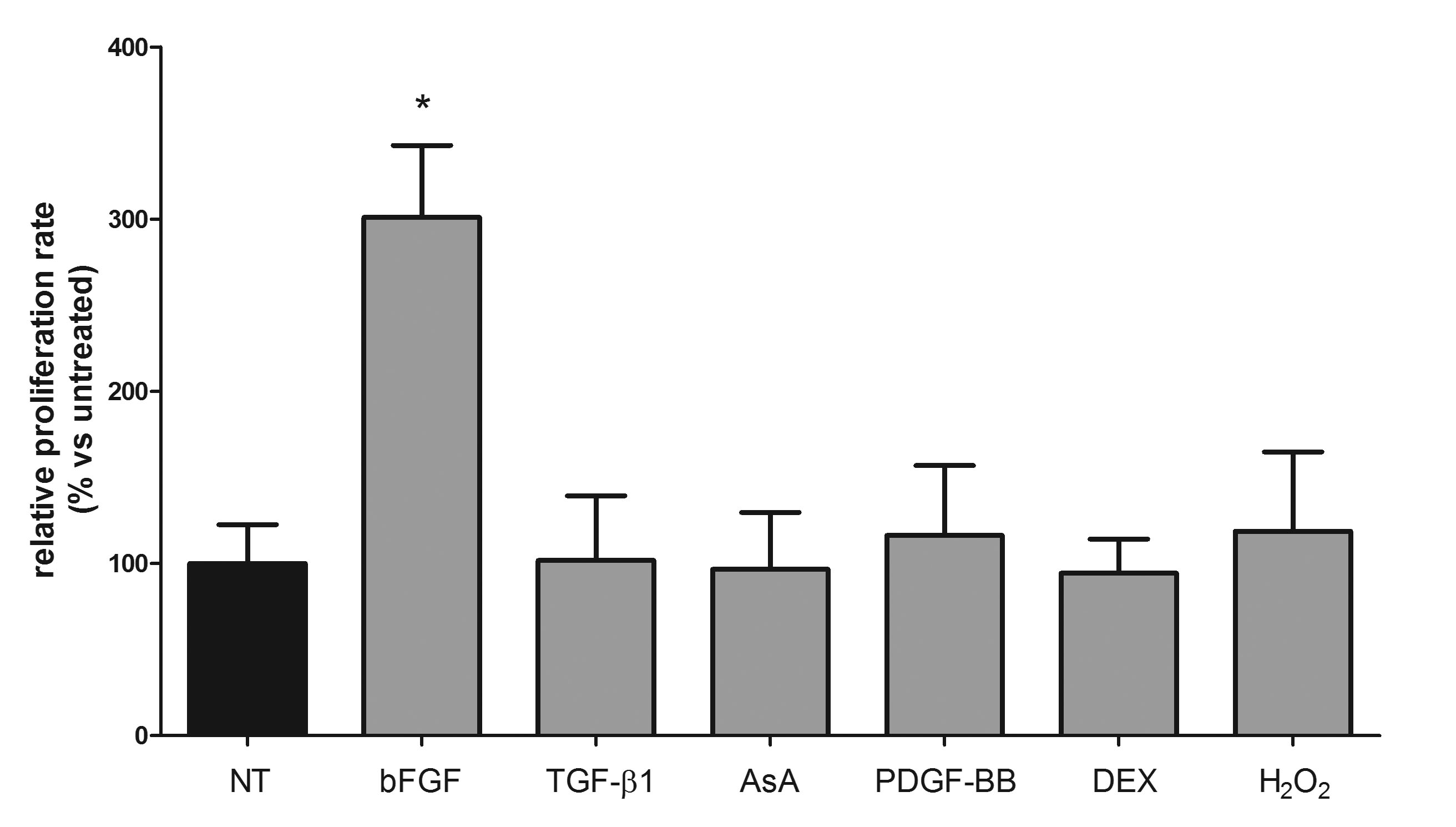
 Figure 5 of
Nuzzi, Mol Vis 2020; 26:818-829.
Figure 5 of
Nuzzi, Mol Vis 2020; 26:818-829.