Figure 4. Immunofluorescent staining. Hyalocytes were stained with phalloidin, showing the cell shape and cytoskeletal changes after
6 days of exposure to the different treatments (doses are indicated in the Methods section). A: Control. B: Basic fibroblast growth factor (bFGF). C: Transforming growth factor beta 1 (TGF-β). D: Platelet-derived growth factor subunit-BB (PDGF-BB). E: Ascorbic acid (AsA). F: Dexamethasone (DEX). G: H2O2.
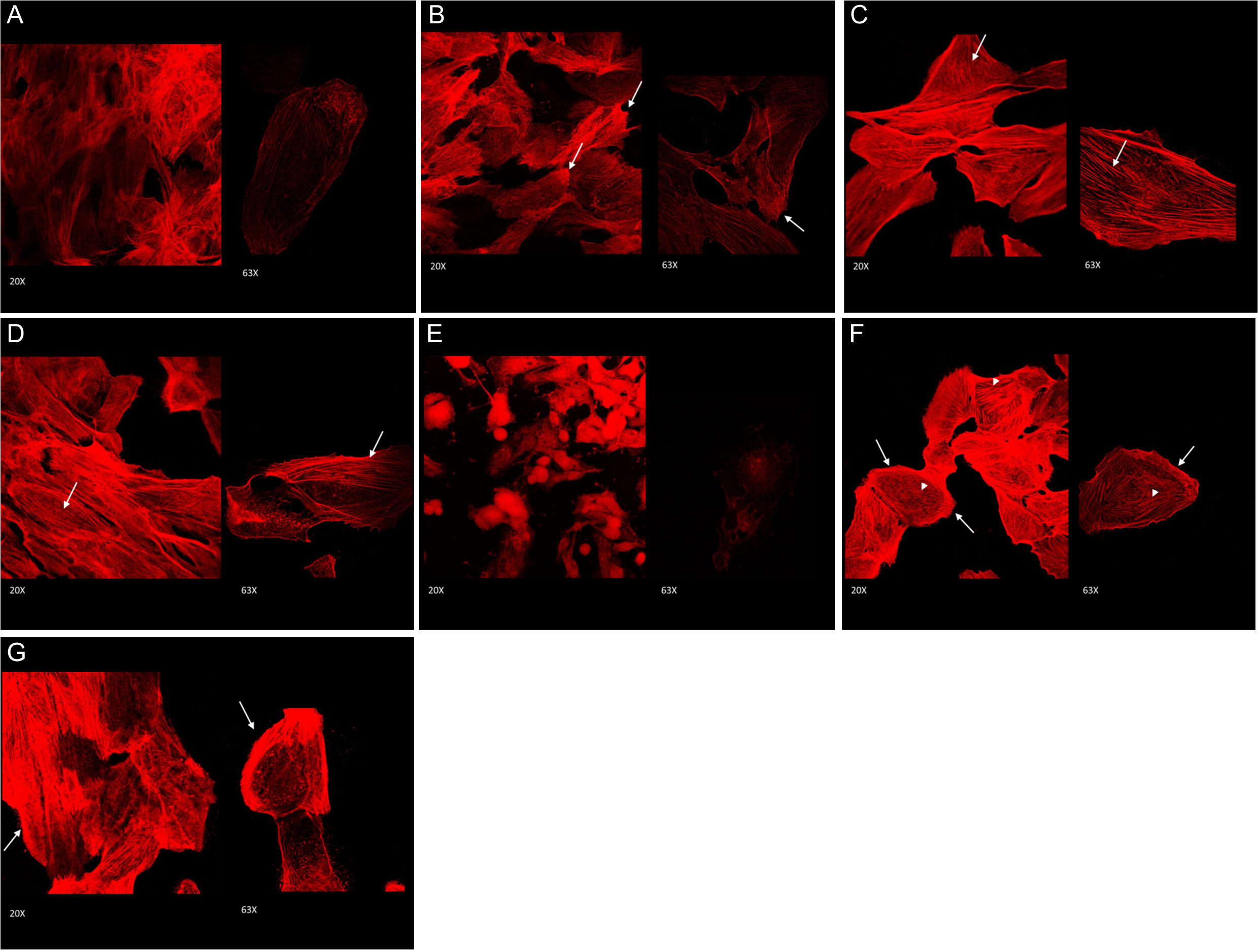
 Figure 4 of
Nuzzi, Mol Vis 2020; 26:818-829.
Figure 4 of
Nuzzi, Mol Vis 2020; 26:818-829.