Figure 2. Subcellular localization of the AQP8 protein in human RPE cells. A: In western blots of the lysates of cell lines from four different donors, the AQP8 protein was found in different bands,
e.g., around 27 and 37 kDa. β-Actin (45 kDa) was used as loading control. B:Double immunolabeling of a cell culture with antibodies against AQP8 (red) and cytochrome c (green). Colocalization of both
immunoreactivities yielded a yellow merge signal. Cell nuclei were stained with 4’,6-diamidine-2-phenylindol (DAPI; blue).
C: Subcellular localization of zonula occludens-1 (ZO-1) immunoreactivity in cultured RPE cells. The arrowheads indicate staining
which lines cell borders. D: Double immunolabeling of cell cultures with antibodies against AQP8 (red) and ZO-1 (green). E:Double immunolabeling of AQP8 (red) and ZO-1 (green) in an RPE monolayer freshly isolated from a donor eye. F: Double immunolabeling of AQP8 (red) and cytochrome c (green) in the freshly isolated RPE monolayer. Scale bars, 20 µm (B‒D) and 10 µm (E, F).
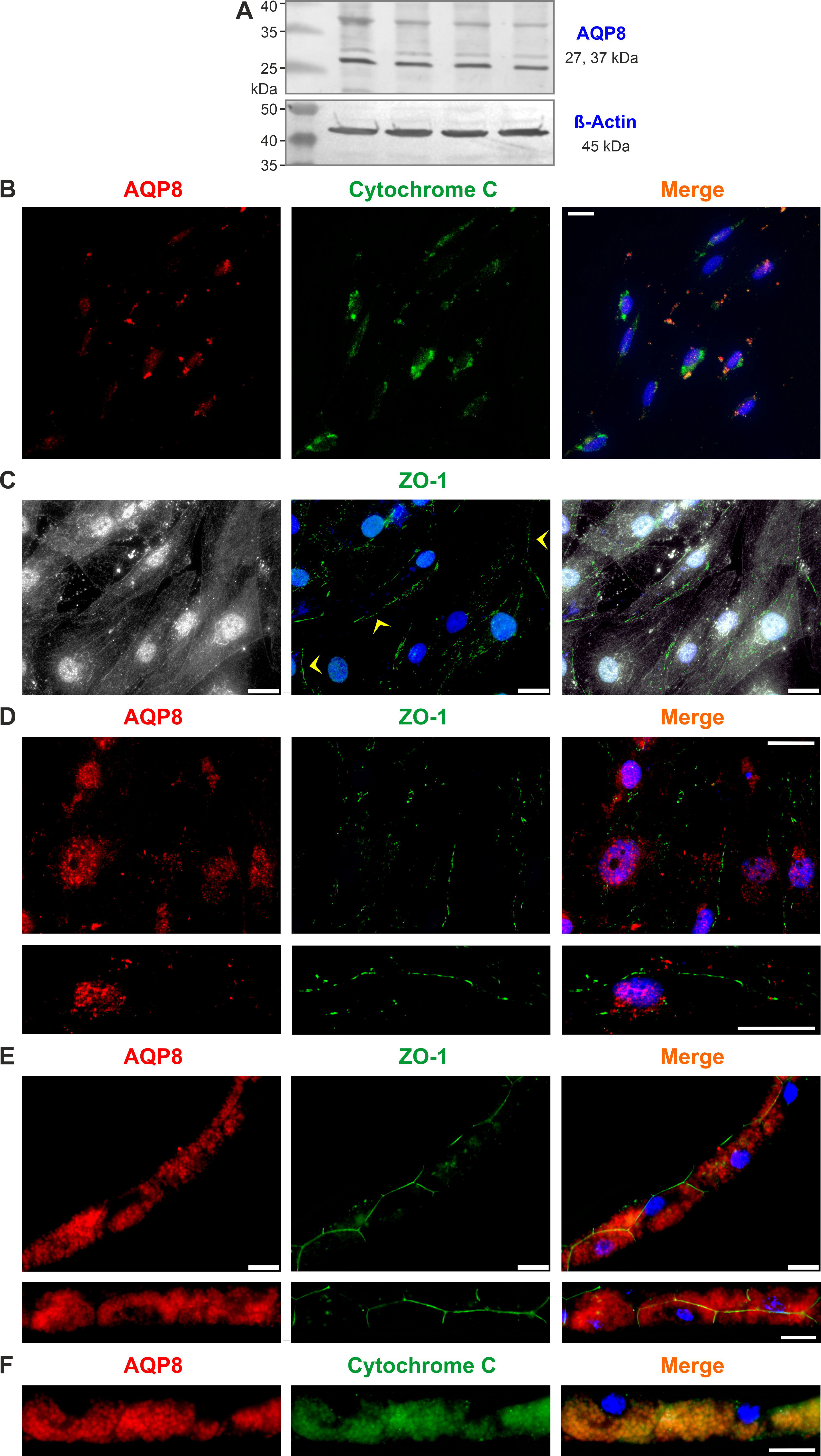
 Figure 2 of
Schnabel, Mol Vis 2020; 26:797-817.
Figure 2 of
Schnabel, Mol Vis 2020; 26:797-817.