Figure 1. Fundus and OCT examination of patient with LHON. A: Evaluation of the fundus shows swelling of the retinal nerve fiber layer (RNFL) in both eyes, hyperemic disc in the right
eye (OD), and diffuse disc pallor in the left eye (OS). B: Optical coherence tomography (OCT) examination displays loss of the ganglion cell layer.
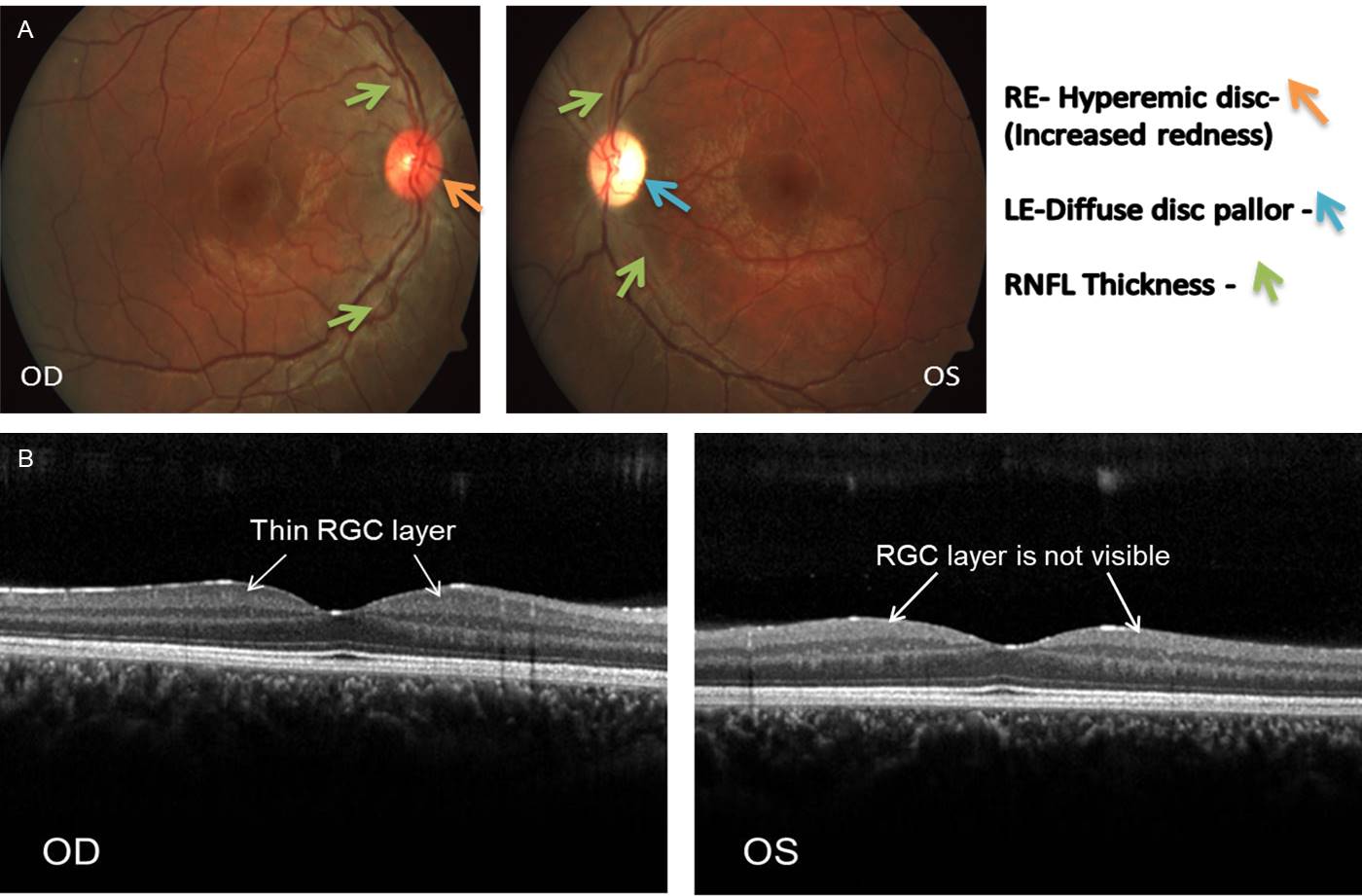
 Figure 1 of
Gowri, Mol Vis 2020; 26:789-796.
Figure 1 of
Gowri, Mol Vis 2020; 26:789-796.