Figure 4. Representative photo series of secreting isolated lacrimal gland duct segments in response to VIP stimulation. The luminal
spaces of the native images (first and third rows) are marked with blue (second and fourth rows). A strong secretory response
could be shown in wild-type (WT) ducts to vasoactive intestinal peptide (VIP) stimulation as the luminal spaces were notably
swollen after treatment. No remarkable changes could be detected in cystic fibrosis transmembrane conductance regulator (CFTR)
knockout (KO) ducts following VIP stimulation.
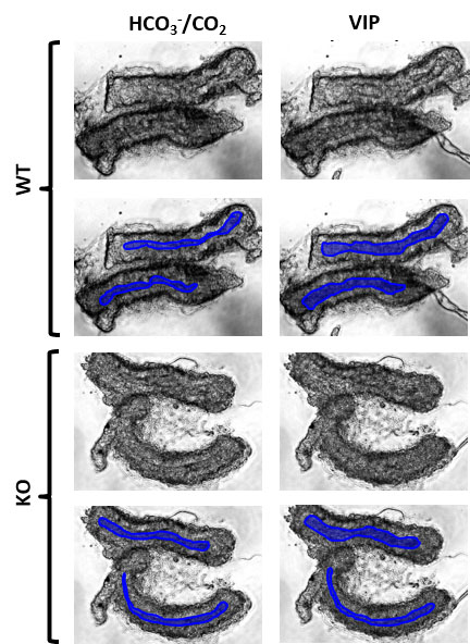
 Figure 4 of
Berczeli, Mol Vis 2020; 26:780-788.
Figure 4 of
Berczeli, Mol Vis 2020; 26:780-788.