Figure 3. Effect of VIP stimulation on ductal fluid secretion in isolated lacrimal gland ducts from WT and CFTR KO mice. A: The wild-type (WT) ducts were exposed either to 100 nM vasoactive intestinal peptide (VIP; filled rhombus) or to no agonist
(empty triangle). B: The cystic fibrosis transmembrane conductance regulator (CFTR) knockout (KO) ducts were exposed either to 100 nM VIP (filled
square) or to 100 nM VIP following 10 µM 1,2-bis(o-aminophenoxy)ethane-N,N,N′,N′-tetra-acetic acid (BAPTA-AM) pretreatment (empty triangle). Changes in relative luminal volume (Vr) are shown. Data were
obtained from six to eight ducts isolated from three different animals in each series and are presented as means ± standard
error of the mean (SEM).
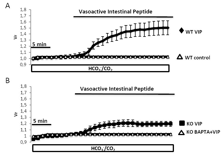
 Figure 3 of
Berczeli, Mol Vis 2020; 26:780-788.
Figure 3 of
Berczeli, Mol Vis 2020; 26:780-788.