Figure 2. Immunofluorescence staining of VPAC2 receptors in lacrimal gland tissues of WT and CFTR mice. VPAC2 was observed on the basolateral
surface of duct (d) and acinar (ac) cells. There were no significant differences between the wild-type (WT) and cystic fibrosis
transmembrane conductance regulator (CFTR) knockout (KO) samples. Hoechst dye was used to stain nuclei blue.
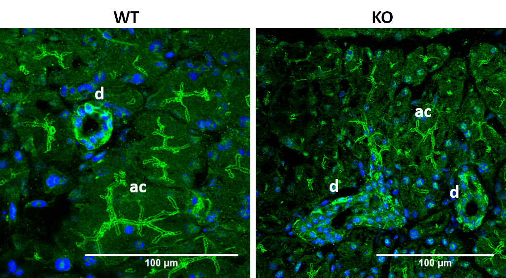
 Figure 2 of
Berczeli, Mol Vis 2020; 26:780-788.
Figure 2 of
Berczeli, Mol Vis 2020; 26:780-788.