Figure 1. Immunofluorescence staining of VPAC1 receptors in lacrimal gland tissues of WT and CFTR KO mice. VPAC1 staining was more intense
in ducts (d) than in acinar (ac) cells. A mosaic pattern was observed in the expression of the receptor proteins in different
ducts. The intensity of the fluorescence varied widely in the investigated duct segments from intense immunofluorescence to
a lack of staining. There were no statistically significant differences between the wild-type (WT) and cystic fibrosis transmembrane
conductance regulator (CFTR) knockout (KO) samples. Hoechst dye was used to stain nuclei blue.
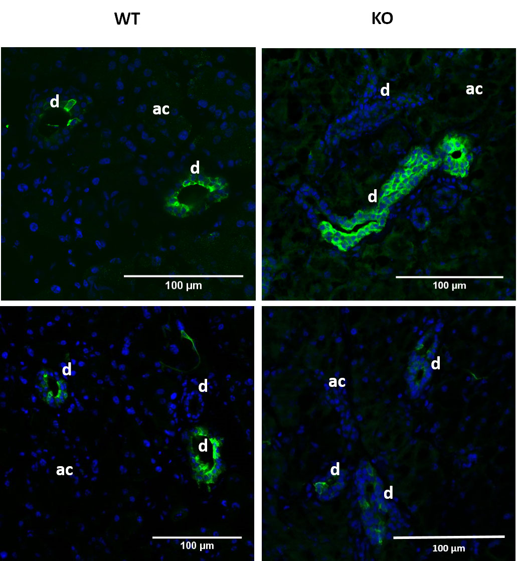
 Figure 1 of
Berczeli, Mol Vis 2020; 26:780-788.
Figure 1 of
Berczeli, Mol Vis 2020; 26:780-788.