Figure 9. Immunoblotting of E2A, α-SMA, and Id3 showing interaction with bHLH E-protein in TGFβ1-induced cellular differentiation in
h-CSFs. A: Representative immunoblots for E2A, alpha smooth muscle actin (α-SMA), Id3, and β-actin. B: Quantification of the corresponding densitometry data of the protein immunoblots. The protein expression was normalized
to β-actin and represented as percentage change (%) compared to the no treatment control (in no transforming growth factor
beta (TGFβ) and Id3 gene transfer groups). Data are mean ± standard deviation (SD, n = 3). *Significant difference from the no treatment control.
This suggests that Id helix–loop–helix (HLH) proteins play a role in the cellular differentiation of human stromal corneal
fibroblasts (h-CSFs) involving TGFβ1.
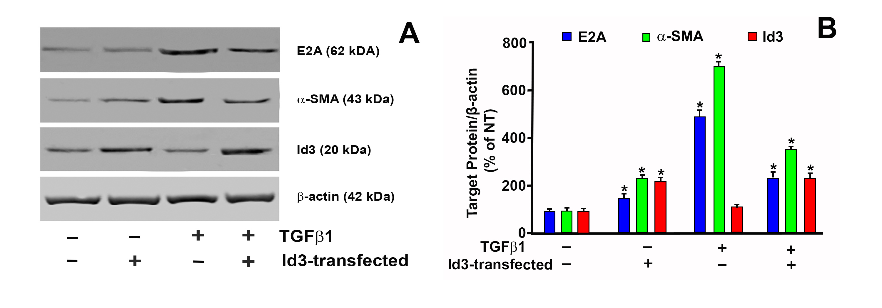
 Figure 9 of
Gupta, Mol Vis 2020; 26:742-756.
Figure 9 of
Gupta, Mol Vis 2020; 26:742-756.