Figure 6. Representative immunofluorescence staining images reveal levels of ⍺-SMA, a myofibroblast marker, and DAPI in h-CSFs. Cultures
with or without Id3 gene transfer were grown in the absence or absence of transforming growth factor beta (TGFβ; 5 ng/ml) for 72 h under serum-free
conditions. A: Normal human stromal corneal fibroblasts (h-CSFs) grown in the absence of TGFβ1. B: Id3-delivered h-CSFs grown in the absence of TGFβ1. C: Normal h-CSFs grown in the presence of TGFβ1. D: Id3-delivered h-CSFs grown in the presence of TGFβ1. TGFβ1 treatments caused a noticeable increase in ⍺-SMA (green) in normal
h-CSFs (C), but this response was prevented by overexpression of the Id3 gene in the h-CSFs (D). Non-TGFβ1-treated cultures mainly exhibited 4′,6-diamidino-2-phenylindole (DAPI) stained nuclei (blue) and the expected
⍺-SMA staining in a few cells (A and B). Magnification bar: 50 µm.
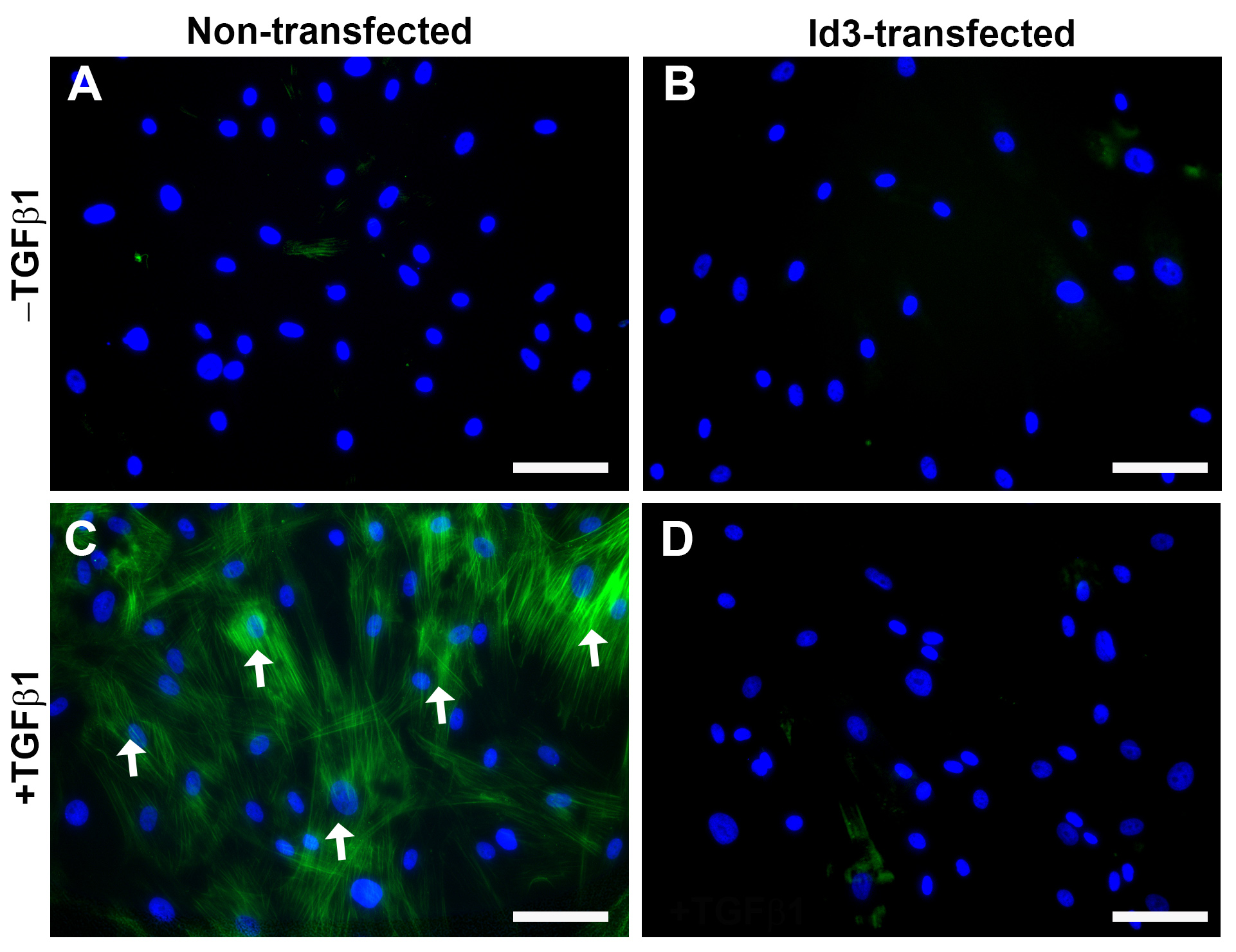
 Figure 6 of
Gupta, Mol Vis 2020; 26:742-756.
Figure 6 of
Gupta, Mol Vis 2020; 26:742-756.