Figure 5. Phase contrast microscopy showing changes in the cellular phenotype of the h-CSFs grown in the presence or absence of TGFβ1
(5 ng/ml) in serum-free conditions with or without Id3 gene transfer. A: Normal human stromal corneal fibroblasts (h-CSFs) grown in the absence of transforming growth factor beta (TGFβ). B: Naked-vector transfected h-CSFs grown in the absence of TGFβ1. C: Id3-transfected h-CSFs grown in the absence of TGFβ1. D: Normal h-CSFs grown in the presence of TGFβ1. E: Naked-vector transfected h-CSFs grown in the presence of TGFβ1. F: Id3-transfected h-CSFs grown in the presence of TGFβ1. Overexpression of the Id3gene in h-CSFs prevented TGFβ-induced cellular differentiation and change to the myofibroblast phenotype (F). Magnification bar: 50 µm.
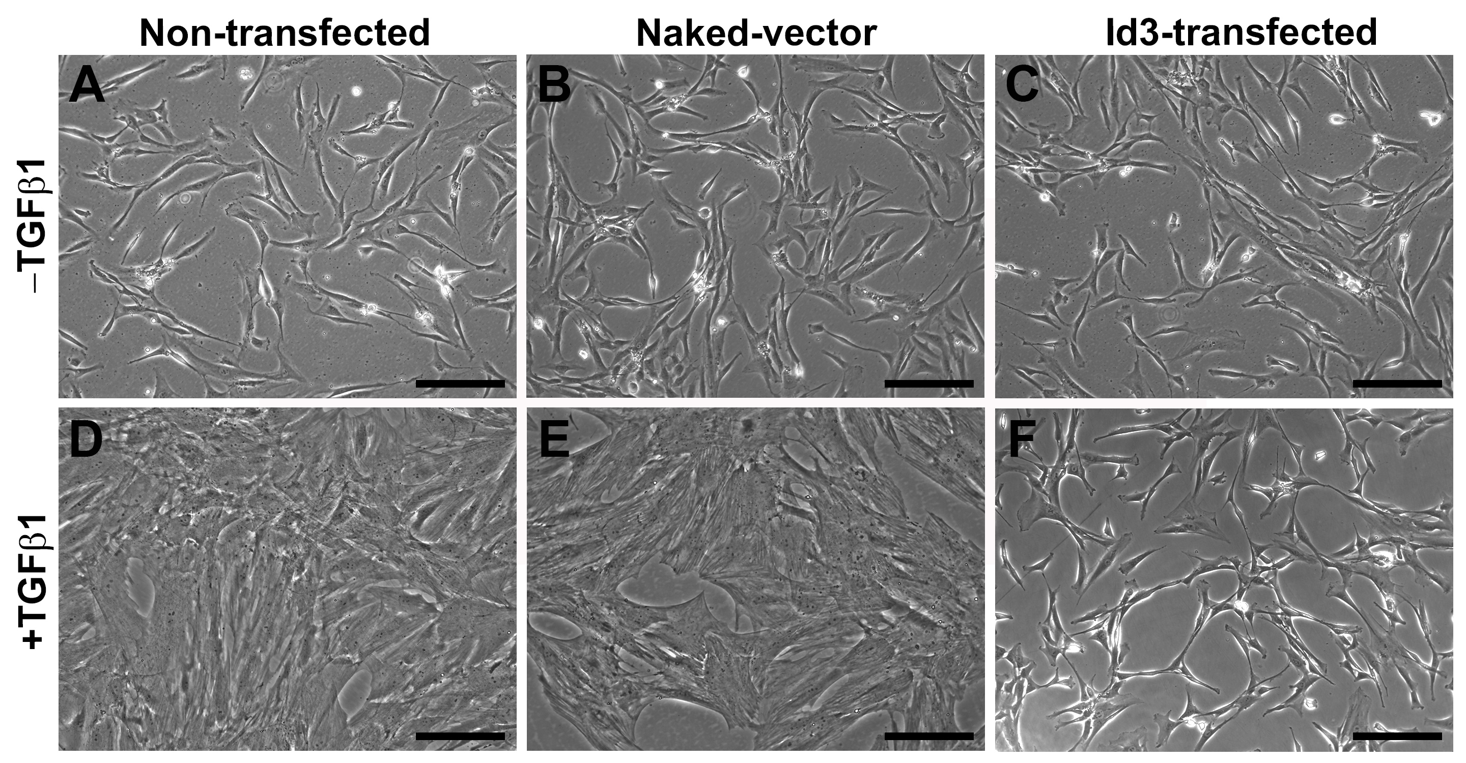
 Figure 5 of
Gupta, Mol Vis 2020; 26:742-756.
Figure 5 of
Gupta, Mol Vis 2020; 26:742-756.