Figure 3. Representative phase contrast microscopy images revealing the phenotype of h-CSFs with and without gene transfer. A: Normal or non-transfected human stromal corneal fibroblasts (h-CSFs). B: Naked-vector transfected h-CSFs. C: Id3-mCherry transfected h-CSFs. No statistically significant differences in cellular phenotype were observed between the Id3-mCherry transfected h-CSFs (C) and the control groups (A, B). Magnification bar: 100 µm.
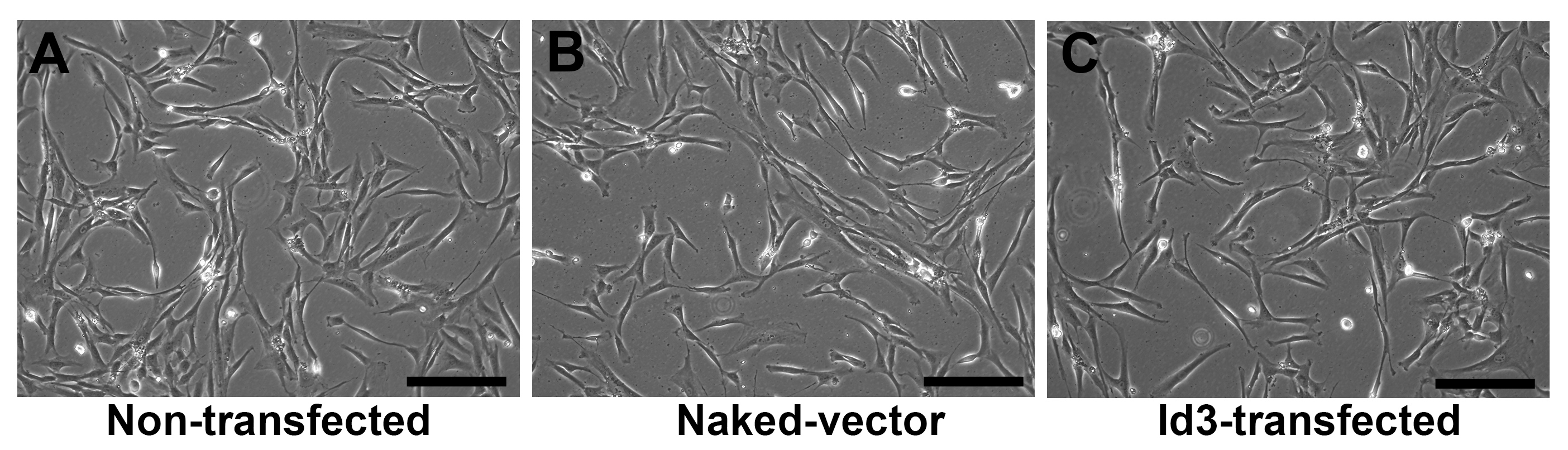
 Figure 3 of
Gupta, Mol Vis 2020; 26:742-756.
Figure 3 of
Gupta, Mol Vis 2020; 26:742-756.