Figure 2. Immunofluorescence images showing the expression of the mCherry in human corneal fibroblast (h-CSFs) in normal and transfected
cells. The Id3-mCherry transfected h-CSFs the successful transfection of Id3 genes (G-I) but non-transfected (A-C) and naked-vector transfected (D-F) cells did not show Id3-mCherry expression as expected. Cells’ nuclei were stained with DAPI (blue color) while Id3-mCherry protein were stained with mCherry (red color) respectively. Magnification bar: 100 μm.
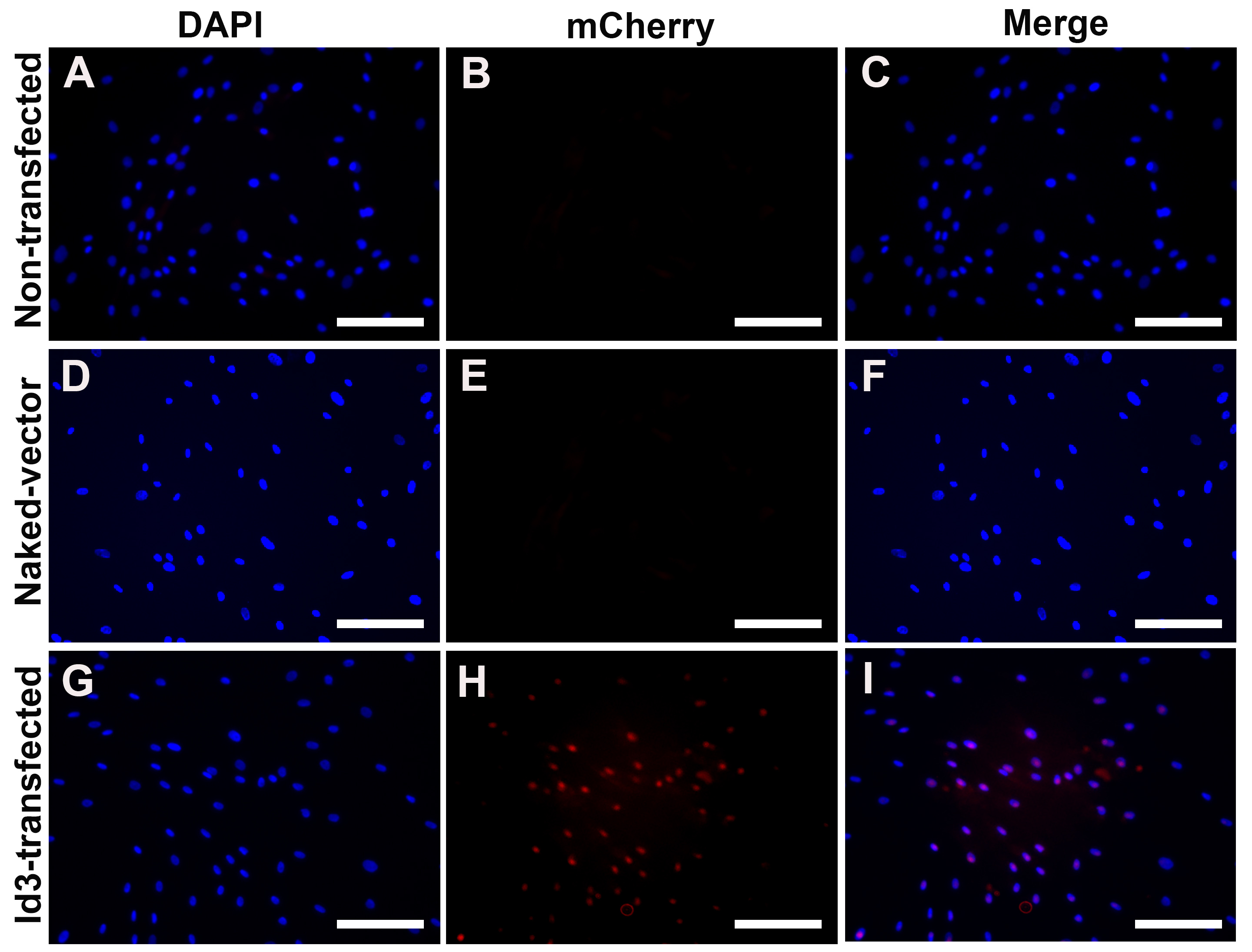
 Figure 2 of
Gupta, Mol Vis 2020; 26:742-756.
Figure 2 of
Gupta, Mol Vis 2020; 26:742-756.