Figure 6. Human arteriovenous crossings. A–C: Images from serial paraffin sections of human retinal arteriovenous crossing sites stained with trichrome. The eye was enucleated
because of a tumor and shows some alteration from the normal retinal aspect. The vessels are within the inner retinal layers
and the vessel wall shows a low density in contrast to pigs. The artery has a slightly thicker wall (stained blue) and a rounded
shape in cross sections as compared to the vein. A–B: The artery is crossing above the vein. C: Two veins that split just before passing the artery are crossing above the latter. D: Immunohistochemical staining of a paraffin section of human retina using antibodies raised against ACTA2 and GFAP. In the
artery (left) and the vein (right), the GFAP staining is surrounding the tunica media (stained by ACTA2), resulting in a complementary
staining pattern that does not overlap. E–H: Immunohistochemical staining of serial paraffin sections of human retinal arteriovenous crossing sites using antibodies
raised against ACTA2 (E and G) and GFAP (F and H). Adjacent sections are compared. The staining is similar to that of porcine crossings. The outer border of the tunica media
is more clearly visible than the trichrome staining. Usually, the undercrossing vein shows a depression at the site where
it is touching the artery. The layers of the vascular wall that are stained by the antibody raised against ACTA2 are touching
but not fusing. E and F: A large vein is undercrossing a large artery in the central retina. G and H: A large vein is undercrossing a small vein in a peripheral part of the retina (compare the thickness of the retina).
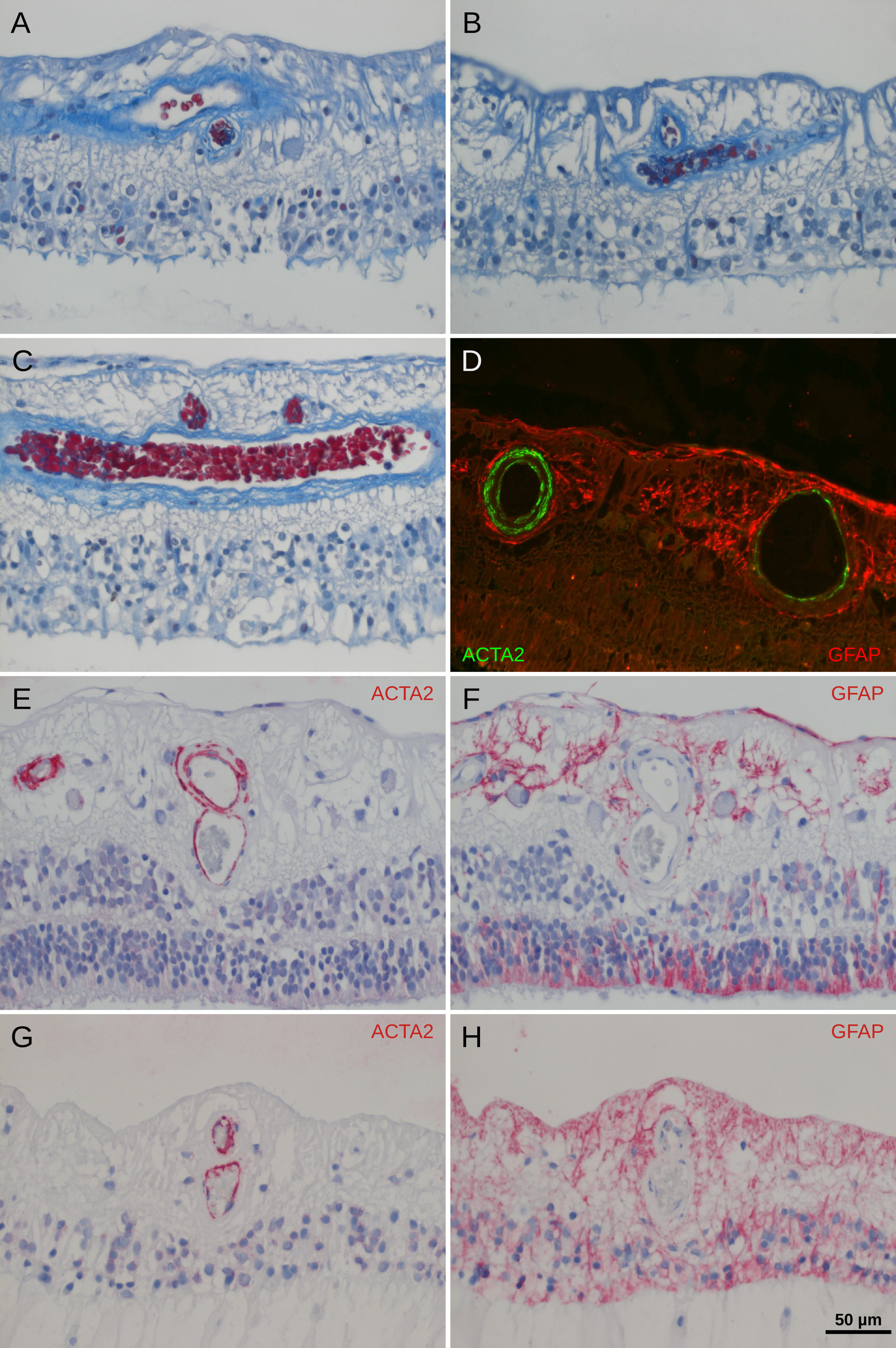
 Figure 6 of
Martin, Mol Vis 2020; 26:731-741.
Figure 6 of
Martin, Mol Vis 2020; 26:731-741.