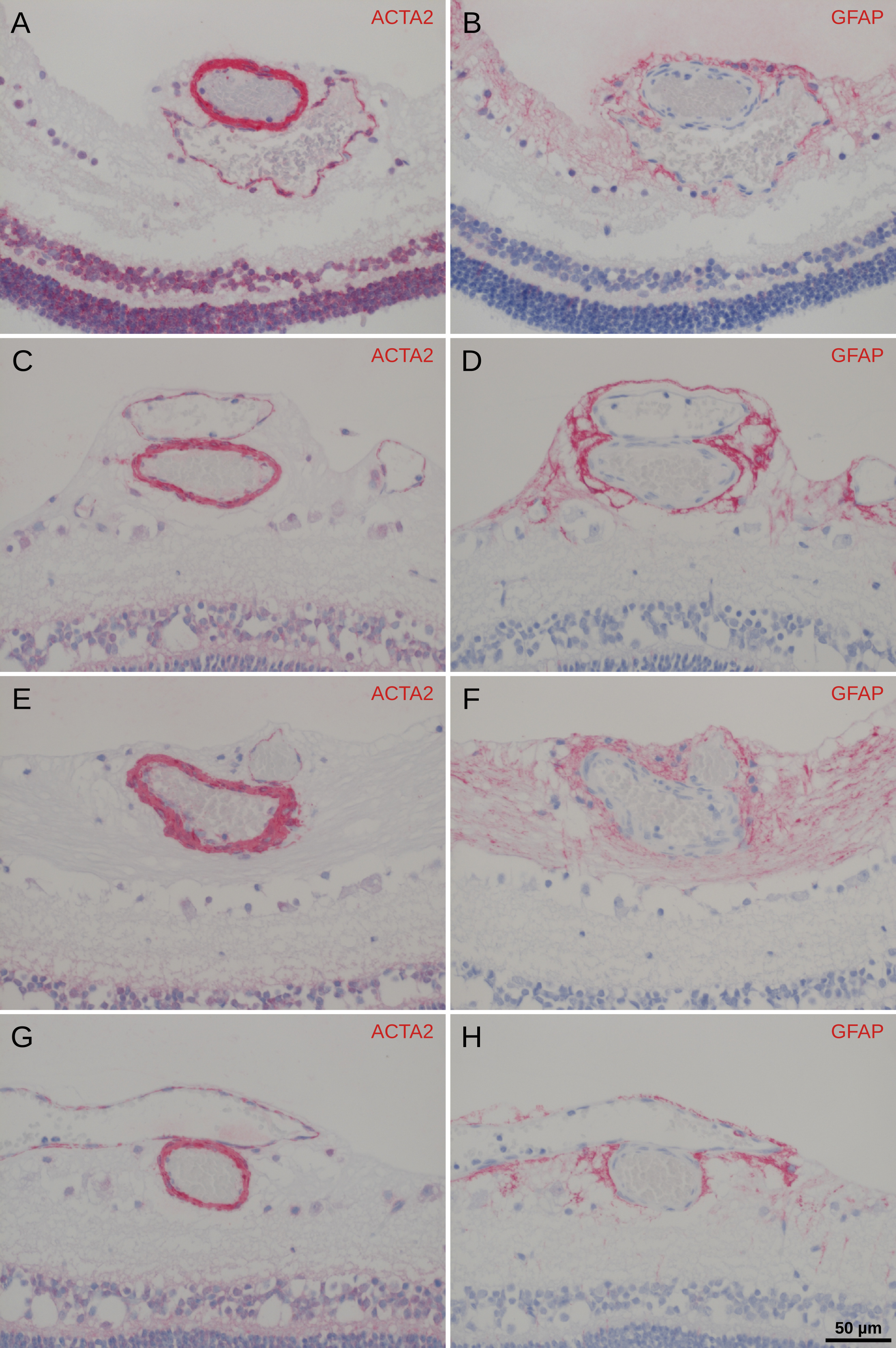
Figure 5. Immunohistochemistry of porcine arteriovenous crossings. Immunohistochemical staining of serial paraffin sections of porcine
retinal arteriovenous crossing sites using antibodies raised against ACTA2 (
A,
C,
E, and
G) and GFAP (
B,
D,
F, and
H). Vicinal sections are compared. While the antibody raised against GFAP stains astrocytes (and some Müller cells) in the
nervous tissue surrounding the vessel, the antibody raised against vascular smooth muscle ACTA2 stains the compact layer of
the vessel wall that is intensely stained by aniline blue in the trichrome staining. The layers of the vascular wall that
are stained by the antibody raised against ACTA2 are touching but not fusing. There is no space left for cells expressing
GFAP between the two vessels at the center of the crossing. Note that the astrocytes show a much higher density around the
vessels.
A and
B show the same arterial overcrossing as in
Figure 4D.
 Figure 5 of
Martin, Mol Vis 2020; 26:731-741.
Figure 5 of
Martin, Mol Vis 2020; 26:731-741.