Figure 3. Rat retinal arteriovenous crossings. Paraffin sections of retinal arteriovenous crossings in rats stained with trichrome.
Arteries are characterized by a slightly thicker wall and a rounded oval cross-section area. These features are prominent
in only some of the serial sections. A–C: The artery (a) is above (toward the vitreous) the vein (v); the vein is cut longitudinally in C. D–H: The vein is crossing over the artery (toward the vitreous). The artery is cut perpendicularly in F, while it is cut obliquely in D, E, G, and H. At all crossing sites, only a small tissue layer separates artery and vein. No obvious compression of the veins was observed.
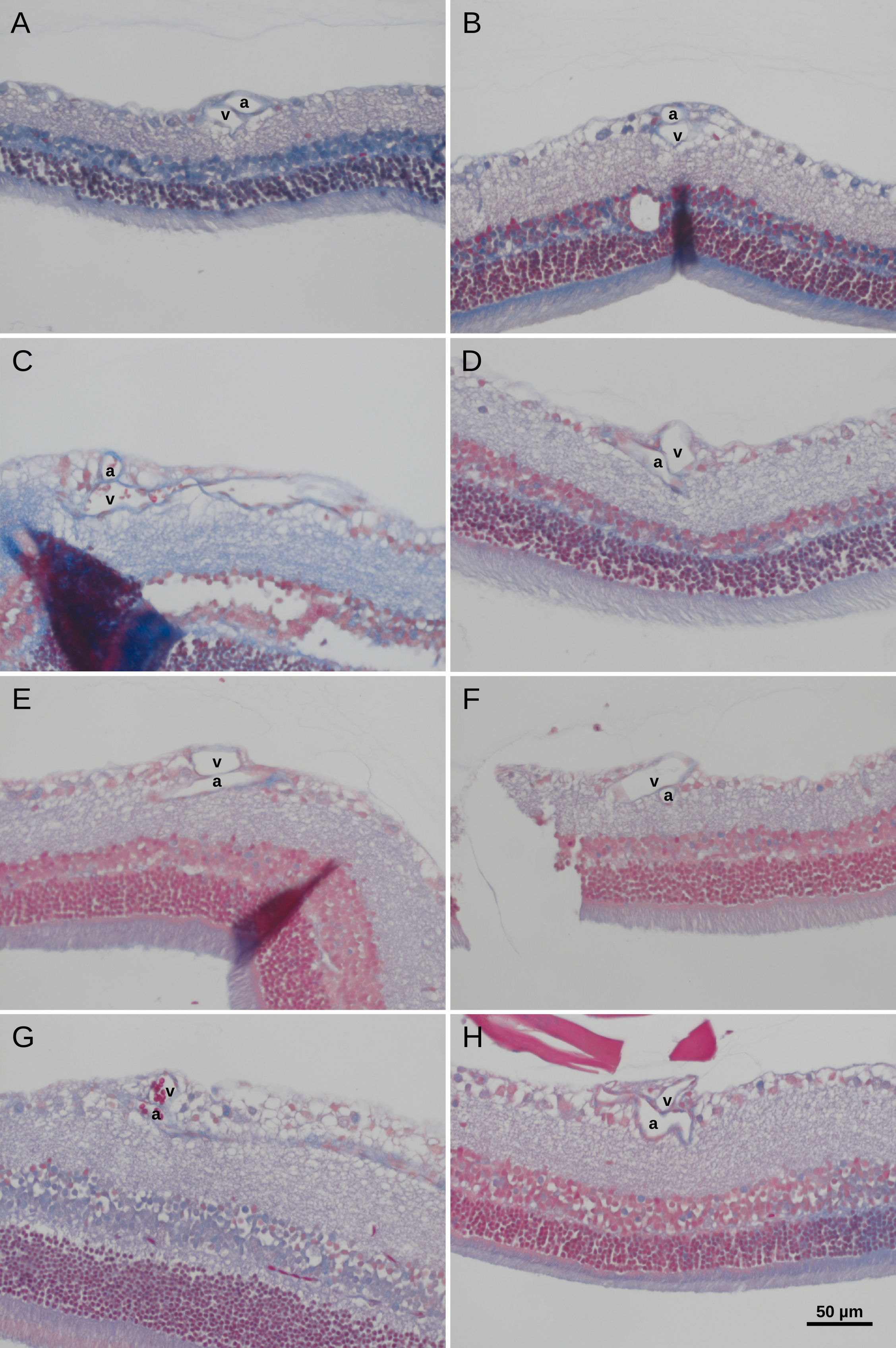
 Figure 3 of
Martin, Mol Vis 2020; 26:731-741.
Figure 3 of
Martin, Mol Vis 2020; 26:731-741.