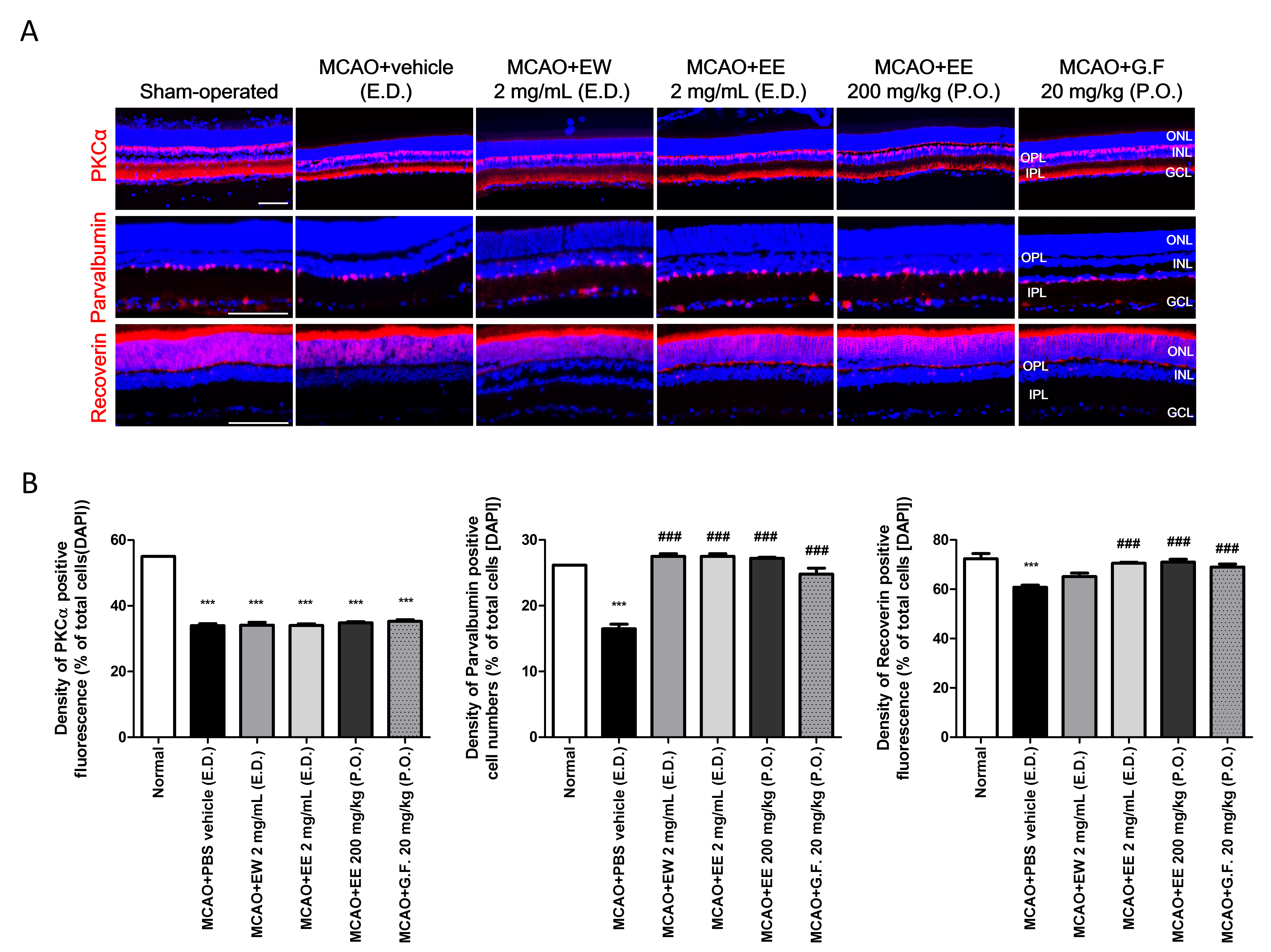
Figure 4. Immunohistochemistry for markers of inner retinal neurons. A: SD rats were treated using topical eye drops (E.D., three times daily) or via oral administration (P.O., once daily) of
KIOM-2015EW, KIOM-2015EE, or Ginexin-F. Five days after treatment, sectioned retinal tissues underwent immunohistochemistry
for the indicated markers (red) and were counterstained with DAPI (blue) after vertical sectioning. Protein kinase C alpha
(PKCα), bipolar; parvalbumin, amacrine; recoverin, photoreceptors. B: Quantification of red fluorescence density was performed using the ImageJ software program. The figures depict the ratio
of red fluorescence to DAPI fluorescence in the same region. All images were acquired at 40× magnification. Scale bar: 100
μm. Data are presented as mean ± standard error of the mean (SEM). ***p<0.001 versus sham-operated, ###p<0.001 versus MCAO+vehicle
(E.D.). MCAO, middle cerebral artery occlusion; RGC, retinal ganglion cell; G.F., Ginexin F; GCL, ganglion cell layer.

 Figure 4 of
Kim, Mol Vis 2020; 26:691-704.
  Figure 4 of
Kim, Mol Vis 2020; 26:691-704.