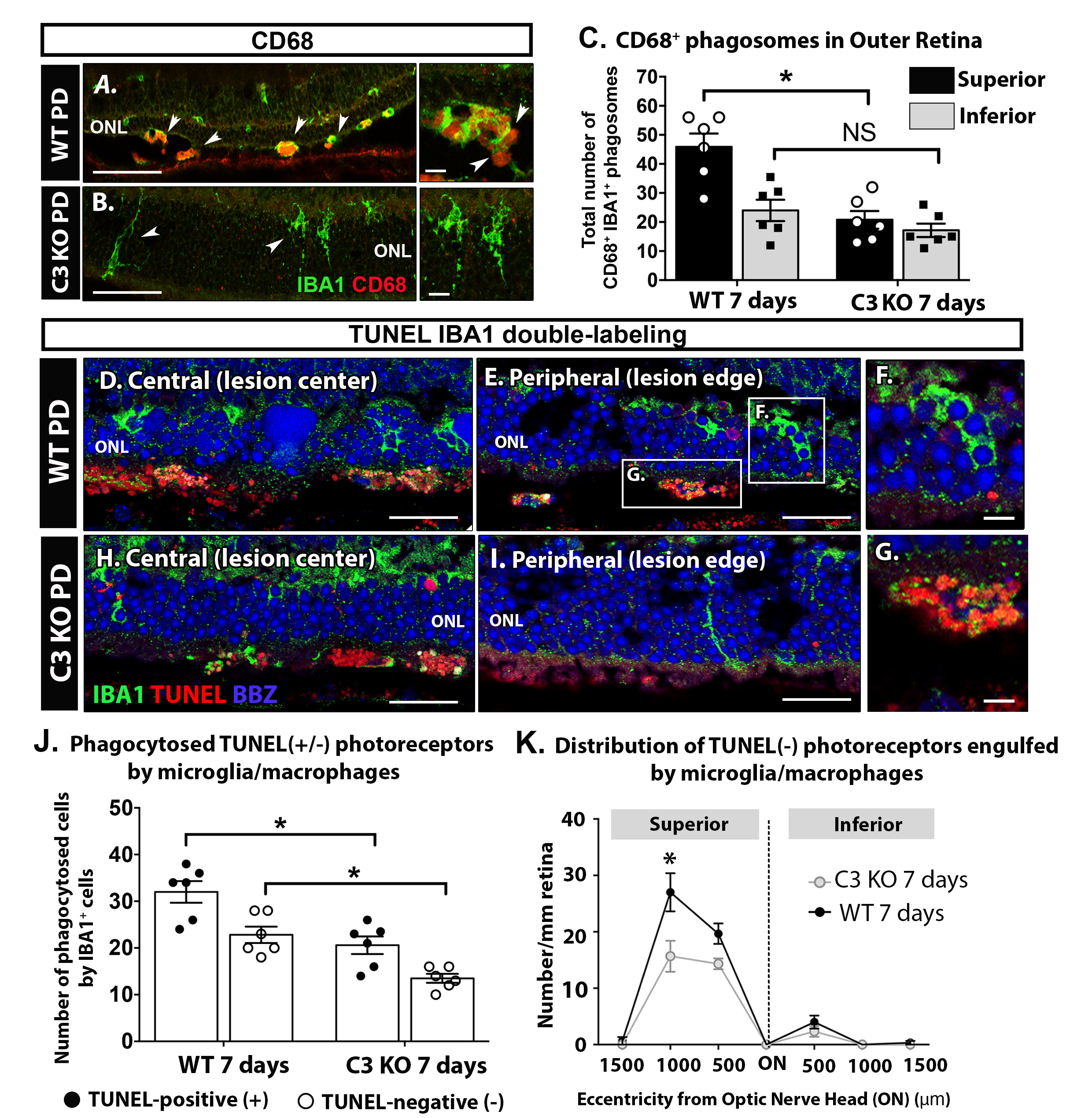
Figure 4. Change in phagocytosis-related properties by macrophages of the outer retina in C3 KO mice after 7 days of PD.
A,
B: Representative vertical sections immunolabeled for CD68/macrosialin (red) and IBA1 (green) to observe outer retinal macrophages
in wild-type (WT) (
A) and C3 knockout (KO) (
B) mice after 7 days of PD.
C: Quantification of CD68-positive/IBA1-positive cells in the outer retina on vertical sections indicates a statistically significant
reduction in these cells in the C3 KO mice compared to the WT mice (p<0.05) following PD.
D–
I: Representative immunolabeling for terminal deoxynucleotidyl transferase dUTP nick end labeling (TUNEL) (red) and IBA1 (green)
to observe changes in the engulfment of TUNEL-positive and -negative photoreceptors between the WT (
D-G) and C3 KO mice (
H-I). Both appeared to be similar in the WT (
D) and C3 KO (
H) groups at the central region of the superior retina which was the lesion center (500 μm eccentricity). In the C3 KO mice
(
I), the engulfment of TUNEL-positive and -negative photoreceptors were observed less frequently at the peripheral region of
the superior retina which was the lesion edge (1,000 μm eccentricity) compared to the WT mice (
E,
F, and
G).
J: Quantification of TUNEL-positive/IBA1-positive (black) or TUNEL-negative/IBA1-positive (white) cells in the outer retina
show that both groups are significantly smaller in the C3 KO cohort compared to the WT cohort (p<0.05) after 7 days of PD.
K: Counts of TUNEL-negative/IBA1-positive cells in 500 μm increments across the vertical meridian reveal a peak in the engulfment
of TUNEL-negative photoreceptors at 1,000 μm eccentricity in the WT mice, which is significantly lower in the C3 KO cohort
(p<0.05). Statistical significance was determined via two-way analysis of variance (ANOVA) with multiple comparisons (n =
5–6 per group/time point; asterisks denote p<0.05). Scale bars represent 50 μm (
A,
B, D,
E,
H,
I) and 10 μm (
F and
G). ONL, outer nuclear layer; ON, optic nerve head; PD, photo-oxidative damage. Panel A, E and G are adapted from [
42].
 Figure 4 of
Jiao, Mol Vis 2020; 26:679-690.
Figure 4 of
Jiao, Mol Vis 2020; 26:679-690.