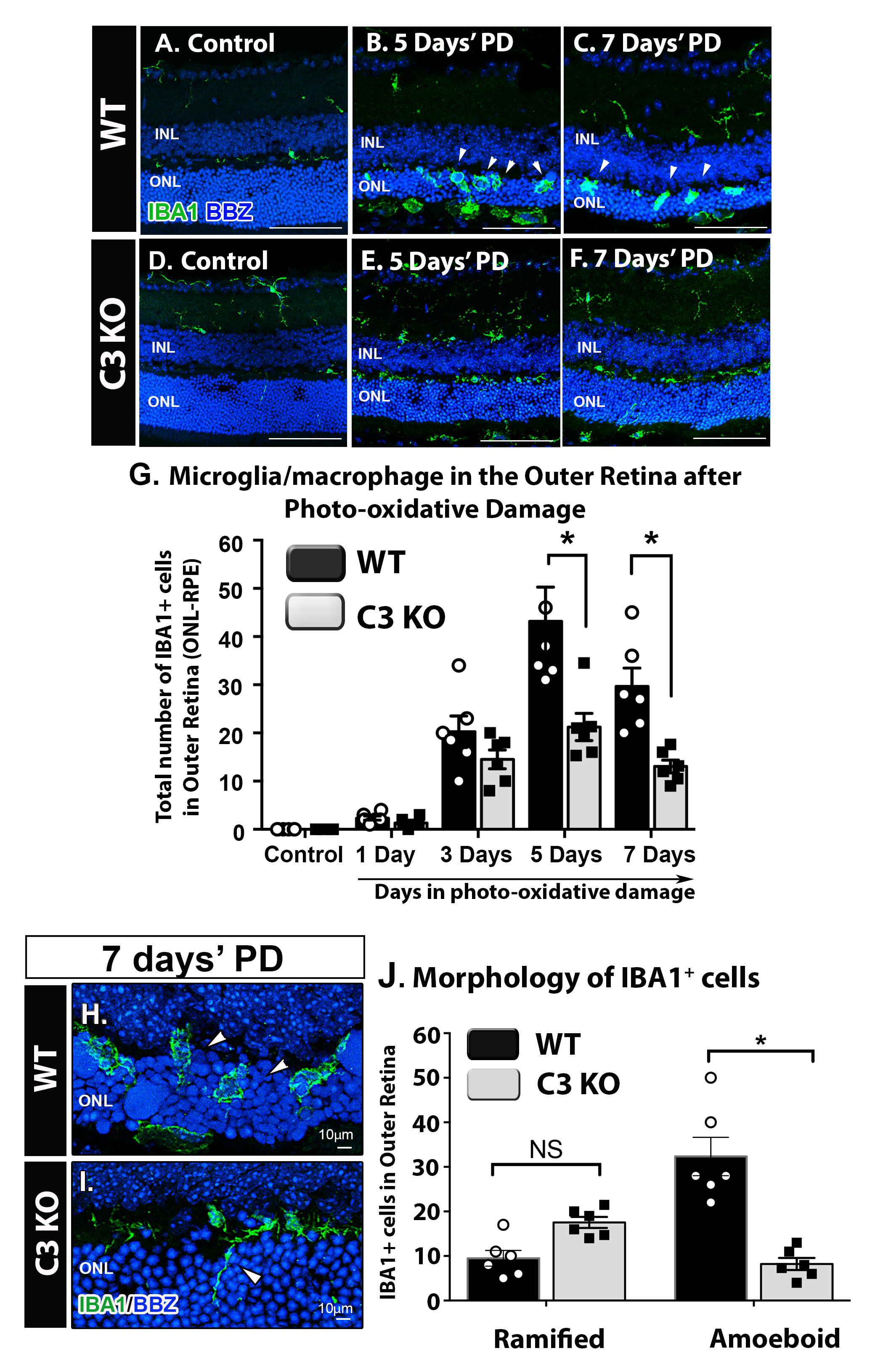
Figure 3. Incursion and morphological change in outer retinal macrophages from WT and C3 KO mice following a time course of PD.
A–
F: Representative retinal cross-sections fluorescently immunolabeled with IBA1 (green) and bisbenzamide (blue), in the 5- and
7-day PD groups. In the dim-reared retinas (
A,
D), IBA1-positive outer retinal macrophages were not present in either the wild-type (WT) or C3 knockout (KO) groups. After
either 5 or 7 days of PD, a substantial incursion of macrophages was observed in the outer retina of the WT mice (
B,
C), which appeared less frequently in the C3 KO mice (
E,
F).
G: Quantification of the average number of IBA1-positive outer retinal macrophages showed a statistically significant reduction
in the C3 KO mice compared to the WT mice at the 5- and 7-day time points (p<0.05) but not at 1 or 3 days (p>0.05).
H–
I: Representative retinal cross-sections fluorescently immunolabeled with IBA1 (green) illustrate differences in the abundance
of ameboid (
H, arrowheads) and ramified (
I, arrowhead) morphology in outer retinal macrophages in the WT and C3 KO mice after 7 days of PD.
J: Quantification of ameboid and ramified IBA1-positive outer retinal macrophages at 7 days of PD revealed a substantial reduction
in ameboid cells in the C3 KO mice compared to the WT mice (p>0.05). There was no statistically significant difference in
the number of ramified IBA1-positive cells in the outer retina (p>0.05). Statistical significance was ascertained with two-way
analysis of variance (ANOVA) with multiple comparisons (n = 6 per group; asterisks denote p<0.05). Scale bars represent 100
μm, unless indicated otherwise. INL, inner nuclear layer; ONL, outer nuclear layer; PD, photo-oxidative damage. Panel A is
adapted from [
42].
 Figure 3 of
Jiao, Mol Vis 2020; 26:679-690.
Figure 3 of
Jiao, Mol Vis 2020; 26:679-690.