Figure 1. Changes in regional photoreceptor degeneration in C3 KO mice after 7 days of PD. A: There was no change in the average number of photoreceptors rows along the vertical meridian in C3 knockout (KO) versus
wild-type (WT) mice retinas (p>0.05). Following PD, the C3 KO mice showed statistically significant preservation of the photoreceptor
rows compared to the WT mice at 1,000 μm eccentricity from the ONL in the superior retina (p<0.05). B, C: Representative toluidine blue staining of retinal cross-sections showed no gross morphological difference between dim-reared
WT (B) and C3 KO (C) groups. D–I: Representative staining of retina cross-sections after 7 days of PD indicate substantial deterioration the ONL in the WT
mice (D), in the central (F, about 500 μm eccentricity) and peripheral (G, about 1,000 μm eccentricity) regions of the superior retina. In the C3 KO mice (E), representative images show severe ONL disruption mainly within the central region (H), while the peripheral region was more spared (I). J–K: Higher magnification examinations of WT mice retinas (J) following PD show the incursion of amoeboid cells in the subretinal space (arrowheads) and disruption of the RPE and Bruch’s
membrane complex which was scarcely documented in the C3 KO mice (K). Statistical significance was determined using two-way analysis of variance (ANOVA; p<0.05, n = 6 per experimental group).
Scale bars represent 100 μm (B, C, D, and E), 50 μm (F, G, H, and I), and 10 μm (J and K). GCL, ganglion cell layer; INL, inner nuclear layer; ONL, outer nuclear layer; RPE, retinal pigment epithelium; PD, photo-oxidative
damage; ON, optic nerve head.
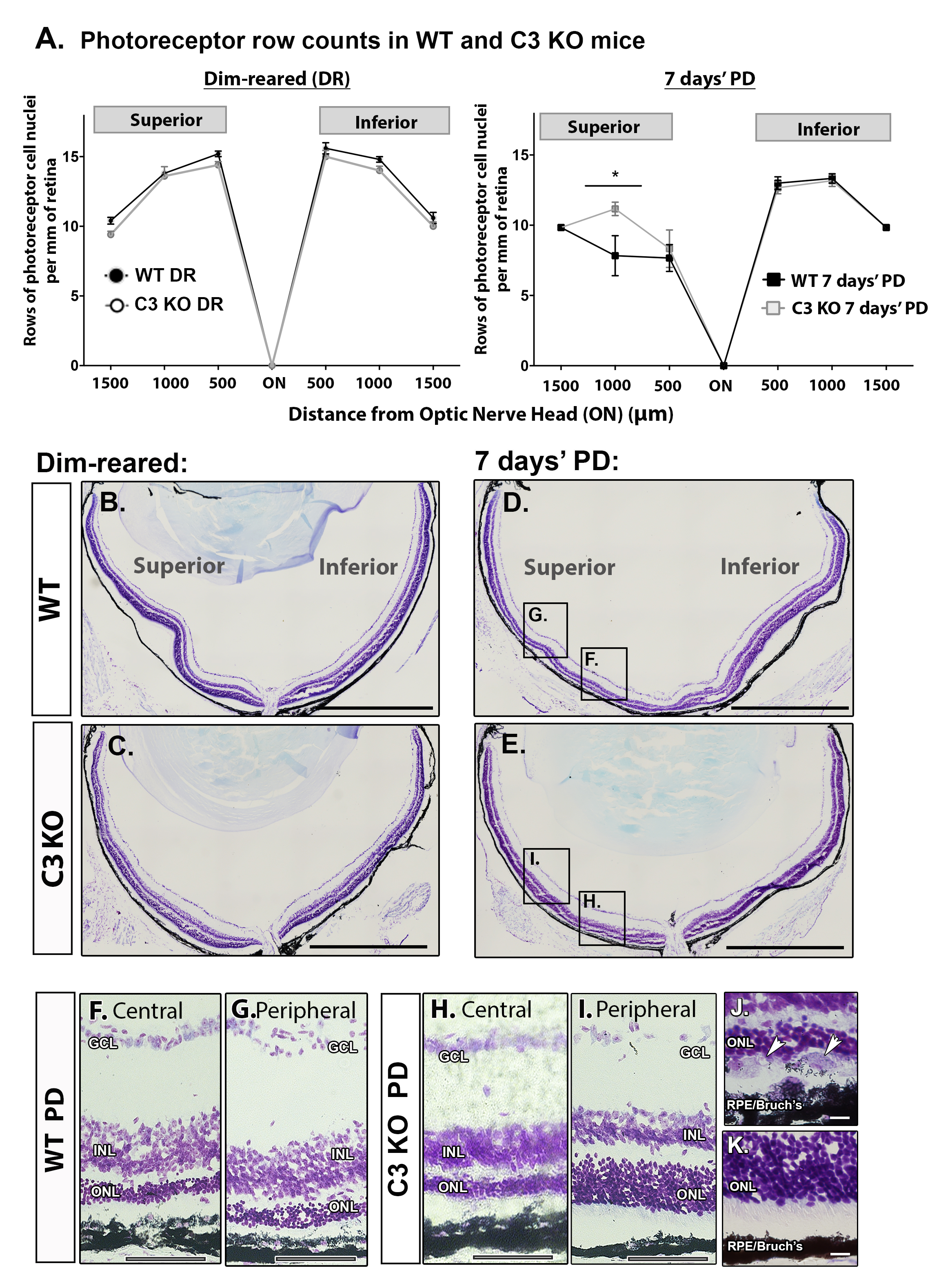
 Figure 1 of
Jiao, Mol Vis 2020; 26:679-690.
Figure 1 of
Jiao, Mol Vis 2020; 26:679-690.