Figure 1. Treatment with tofacitinib inhibits the development of EAU. Groups of B10.A mice were immunized with interphotoreceptor retinoid-binding
protein (IRBP) and treated twice daily with tofacitinib or the vehicle. On day 14, the mice were euthanized, and their eyes
were analyzed histologically for pathological changes typical of experimental autoimmune uveitis (EAU). Panels A-D show minor pathological changes in the eye of a tofacitinib-treated mouse, scored at 0.5+. The changes include accumulation
of inflammatory cells at the limbal (panel B) and optic nerve head (panel C) areas (the two entry sites for the inflammation-inducing cells), as well as small numbers of inflammatory cells in the vitreous
(panel D). Panels E-H are sections of an eye of a control mouse scored at 1.5+. Panel F shows infiltration at the limbal area, as well as typical perivascular accumulation of inflammatory cells (black arrow) and
photoreceptors loss (white arrow). Panel G shows intense infiltration of inflammatory cells at the optic nerve head; panel H shows the typical retinal folding and infiltration of inflammatory cells throughout the retina. I: Summary of the histological data collected in five repeated experiments, with three to five mice per group. Treatment with
tofacitinib significantly inhibited the development of disease. J: The actual numbers of CD4+ and CD8+ T cells, as well as NK cells (CD3-NK1.1+) in the eyes of the immunized mice treated with vehicle or tofacitinib. The means ± standard error of the mean (SEM) from
individual mice from a representative experiment; similar findings were made in three additional experiments (n = 5/group)
* p<0.05. Student t test.
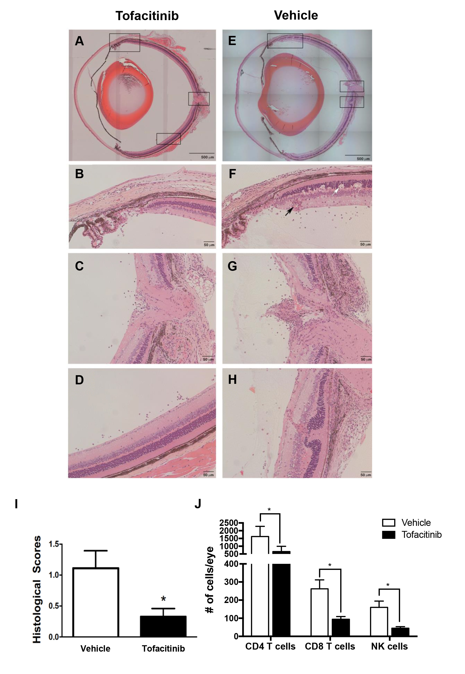
 Figure 1 of
Bing, Mol Vis 2020; 26:641-651.
Figure 1 of
Bing, Mol Vis 2020; 26:641-651.