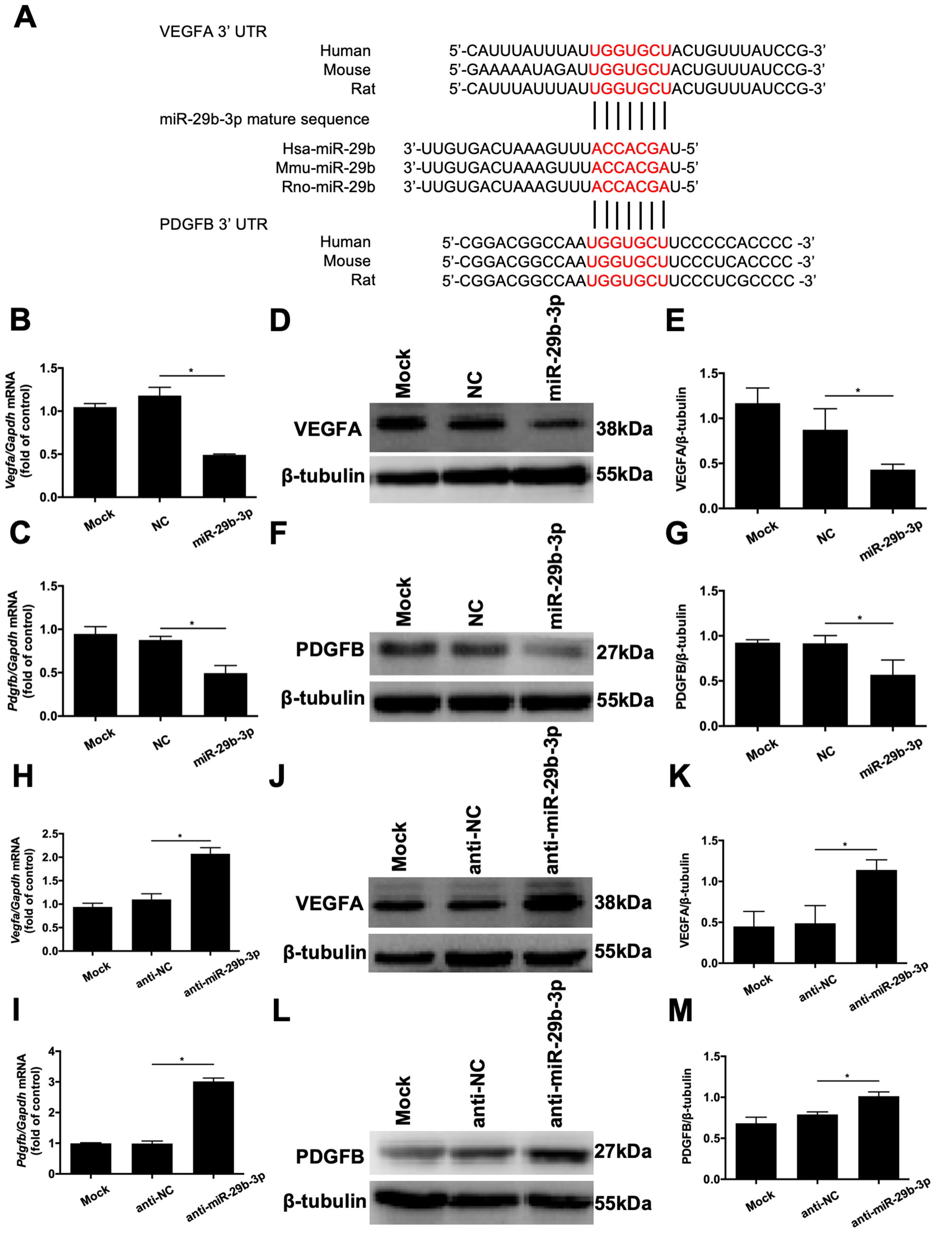
Figure 6. VEGFA and PDGFB are potential targets of microRNA (miR)-29b-3p. A: Bioinformatics analysis revealed that the miR-29b-3p recognition sites (shown in red) on the 3′-untranslated regions of
vascular endothelial growth factor A (VEGFA) and platelet-derived growth factor B (PDGFB) are highly conserved among rat (rno-),
human (hsa-), and mouse (mmu-) genes. B, C: Real-time quantitative PCR analysis revealed that transfection of retinal microvascular endothelial cells (RMECs) with miR-29b-3p
statistically significantly inhibited the mRNA expression of VEGFA and PDGFB compared with that of the negative control (NC)
group at 48 h after transfection. D–G: At 72 h after transfection, western blotting revealed that miR-29b-3p decreased the protein expression levels of VEGFA and
PDGFB compared with those of the NC group. H, I: At 48 h after transfection, real-time quantitative PCR revealed that anti-miR-29b-3p statistically significantly increased
the mRNA expression levels of VEGFA and PDGFB in RMECs compared with those of the anti-NC group. J–M: At 72 h after transfection, western blotting revealed that anti-miR-29b-3p increased the protein expression levels of VEGFA
and PDGFB in RMECs compared with those of the anti-NC group. Glyceraldehyde 3-phosphate dehydrogenase (GAPDH) and β-tubulin
were used as the internal controls for quantitative real-time PCR and western blotting, respectively. n=3 per group. *p<0.05.

 Figure 6 of
Tang, Mol Vis 2020; 26:64-75.
  Figure 6 of
Tang, Mol Vis 2020; 26:64-75.