Figure 5. Effects of anti-microRNA (miR)-29b-3p on the migration and angiogenesis of RMECs. A: Representative images of wound scratch assays after transfecting retinal microvascular endothelial cells (RMECs) with 100
nmol/l of anti-miR-29b-3p or negative control inhibitor (anti-NC). Images were taken at 0 and 6 h after the wound layer was
scratched with a pipette tip. Scale bar: 500 µm. B: Statistical analysis of wound healing rates in each group revealed that transfection with anti-miR-29b-3p enhanced RMEC
migration compared with that of the anti-NC group. C: Representative images of tube formation assays at 6 h after seeding RMECs transfected with 100 nmol/l of anti-miR-29b-3p
or anti-NC on Matrigel. Scale bar: 100 µm. D: Statistical analysis of the relative change in tube length revealed that transfection with anti-miR-29b-3p statistically
significantly increased RMEC angiogenesis compared with that of the anti-NC group. The tube length was quantified by calculating
the cumulative length of the tubes in each image, and the relative change was compared against the mock-transfected group.
n=3 per group. *p<0.05.
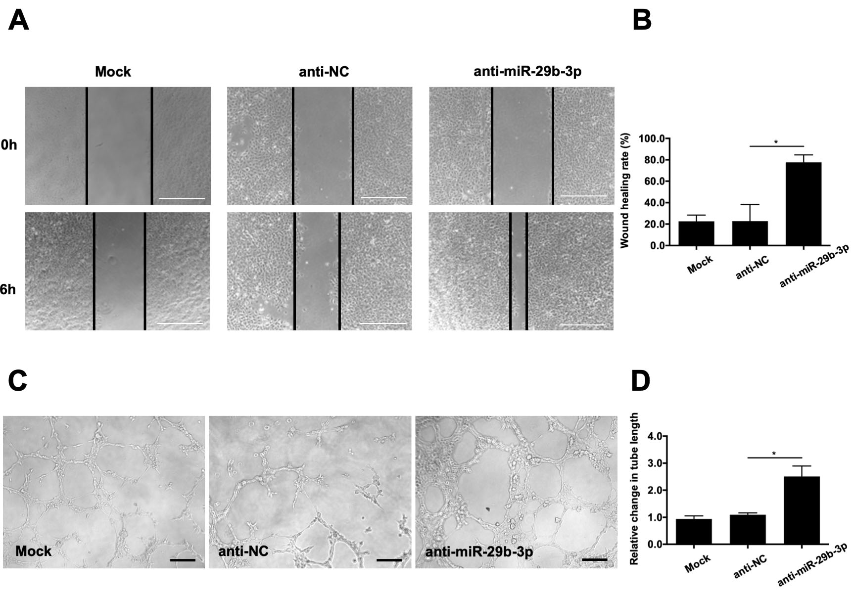
 Figure 5 of
Tang, Mol Vis 2020; 26:64-75.
Figure 5 of
Tang, Mol Vis 2020; 26:64-75.