Figure 4. Effects of anti-microRNA (miR)-29b-3p on the proliferation of RMECs. A: Retinal microvascular endothelial cells (RMECs) were transfected with 100 nmol/l of anti-miR-29b-3p or negative control
inhibitor (anti-NC). Quantitative real-time PCR confirmed that transfection with anti-miR-29b-3p decreased the expression
level of miR-29b-3p compared with that of the anti-NC group. B: At 48 h after transfection, cell viability was assessed using CCK-8 assays, and relative cell viability was calculated against
mock-transfected cells. Transfection with anti-miR-29b-3p statistically significantly increased the viability of RMECs compared
with that of the anti-NC group. C, D: At 48 h after transfection, immunofluorescent staining showed that transfection with anti-miR-29b-3p increased the proportion
of Ki67-labeled cells compared with that of the anti-NC group. The relative proliferation rate of RMECs determined by Ki67
staining was calculated against mock-transfected cells. E, F: At 72 h after transfection, western blotting showed that the protein expression of proliferating cell nuclear antigen (PCNA)
was statistically significantly increased by anti-miR-29b-3p compared with that of the NC group. G–J: At 72 h after transfection, western botting showed that the protein expression levels of cyclin A2, cyclin D1, and cyclin
E1 were statistically significantly upregulated by anti-miR-29b-3p compared with those of the anti-NC group. U6 and β-tubulin
were used as the internal controls for miR-29b-3p expression and western blotting, respectively. n=3 per group. *p<0.05.
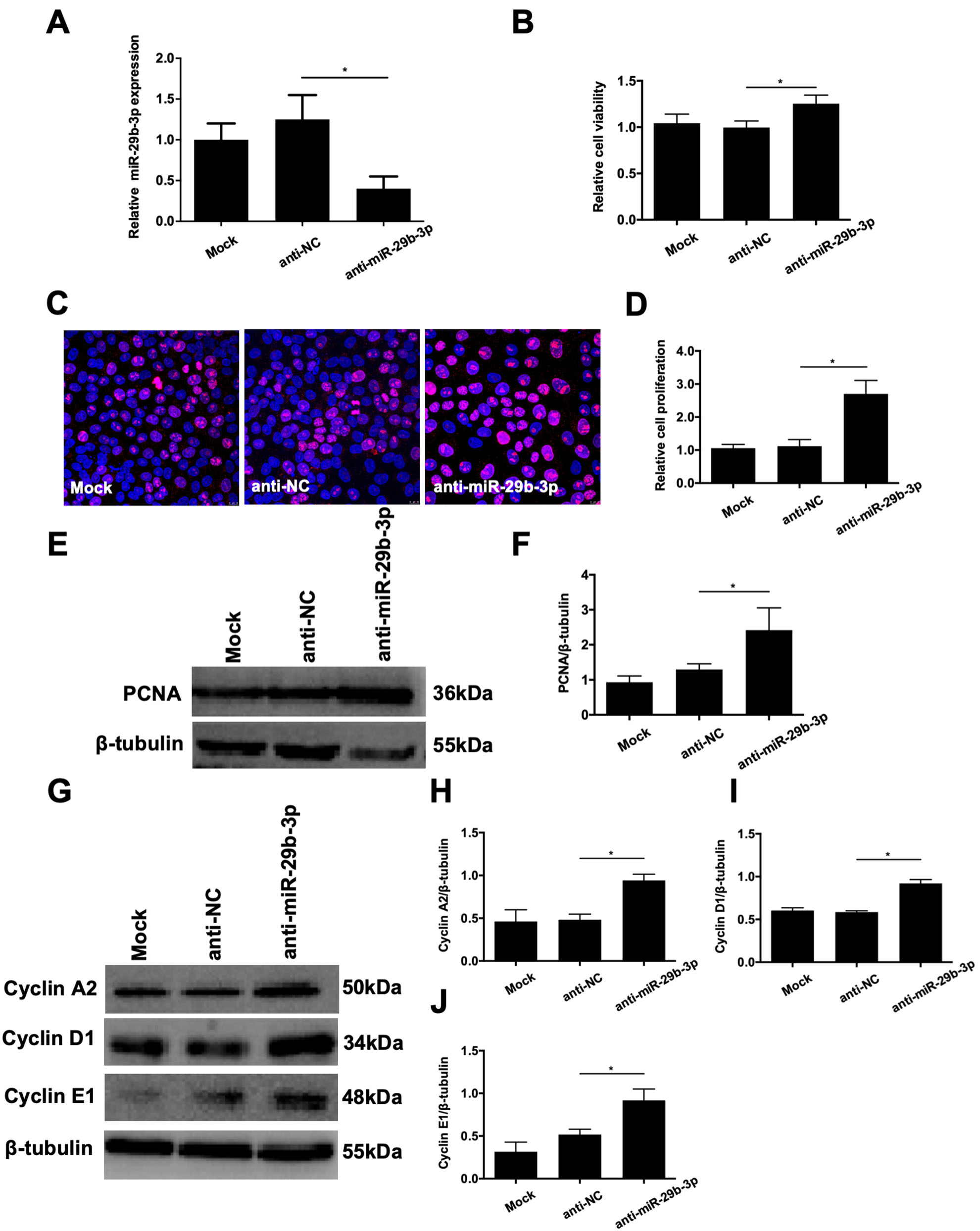
 Figure 4 of
Tang, Mol Vis 2020; 26:64-75.
Figure 4 of
Tang, Mol Vis 2020; 26:64-75.