Figure 3. Effects of microRNA (miR)-29b-3p on the migration and angiogenesis of RMECs. A: Representative images of wound scratch assays after transfecting retinal microvascular endothelial cells (RMECs) with 100
nmol/l of miR-29b-3p-mimic or negative control (NC) mimic. Images were taken at 0 and 6 h after the cell layer was scratched
with a pipette tip. Scale bar: 500 µm. B: Statistical analysis of the wound healing rates revealed that transfection with miR-29b-3p-mimic statistically significantly
inhibited RMEC migration compared that of with the NC group. C: Representative images of tube formation assays at 6 h after seeding RMECs transfected with 100 nmol/l of miR-29b-3p or NC
on Matrigel. Scale bar: 100 µm. D: Statistical analysis of the relative change in tube length revealed that transection with miR-29b-3p-mimic statistically
significantly inhibited tube formation in RMECs compared with that of the NC group. Tube length was quantified by calculating
the cumulative length of the tubes in each image, and the relative change was compared against the mock-transfected group.
n=3 per group. *p<0.05.
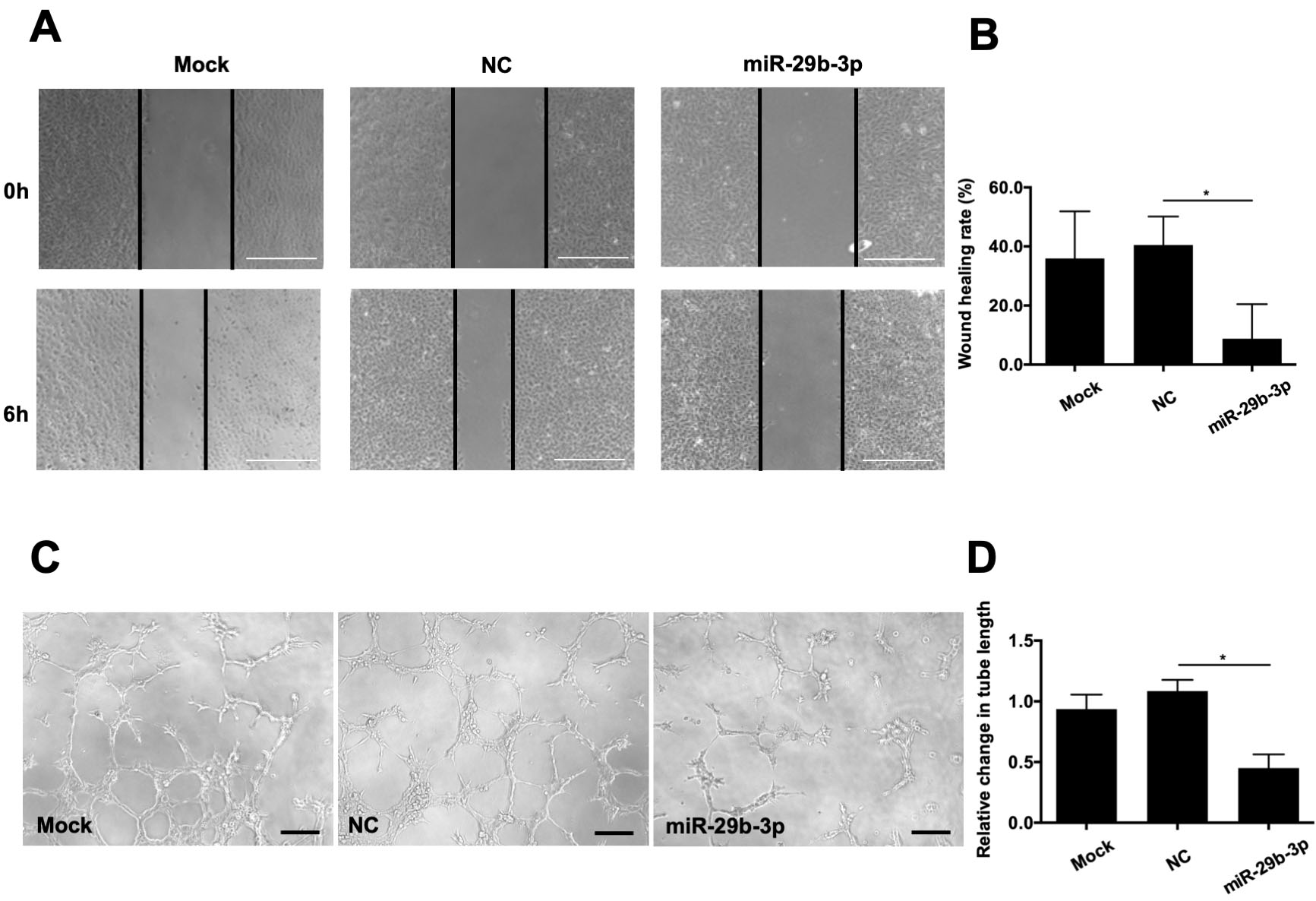
 Figure 3 of
Tang, Mol Vis 2020; 26:64-75.
Figure 3 of
Tang, Mol Vis 2020; 26:64-75.