Figure 1. Characterization of rat primary RMECs. Light microscopic bright-field (BF) images showing the fusiform shape of retinal microvascular
endothelial cells (RMECs). Immunofluorescence (IF) of CD31-labeled RMECs. The endothelial cell marker CD31 was primarily expressed
on the cell membrane. 4′,6-diamidino-2-phenylindole (DAPI) was used to label cell nuclei. Scar bar: 50 µm.
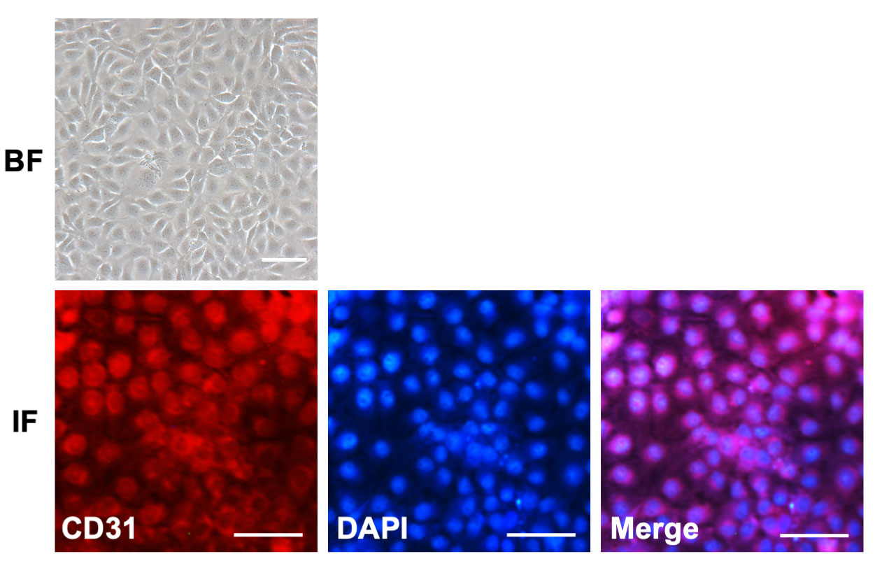
 Figure 1 of
Tang, Mol Vis 2020; 26:64-75.
Figure 1 of
Tang, Mol Vis 2020; 26:64-75.