Figure 4. TRPV1 axons terminate in a simple or ramifying morphology. A: The top panel shows axon terminals expressing TRPV1 and CGRP at the surface of the cornea. The orthogonal view below is
generated by projecting the z-series below in the x-plane. The arrowhead to the right of the y,z panel below indicates the location of the section above
containing ramifying terminals. B: Cross section of the cornea epithelium (12 µm cryosection), sub-basal to surface, demonstrates the colocalization of TRPV1
and CGRP in an axon terminal with simple morphology. As in A, the green arrowhead indicates the surface epithelium, the blue bar indicates the epithelial layers, and the red arrowhead
points to the sub-basal epithelium. (A, B) Scale bar: 5 µm.
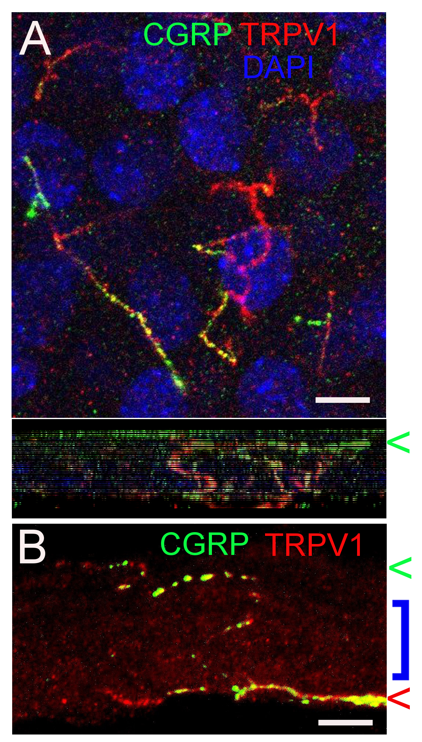
 Figure 4 of
Schecterson, Mol Vis 2020; 26:576-587.
Figure 4 of
Schecterson, Mol Vis 2020; 26:576-587.